


2018 Annual Report
Cover captions, from top to bottom:
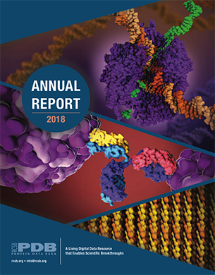
RNA polymerase is a complex enzyme at the heart of transcription. During this process, the enzyme unwinds the DNA double helix and uses one strand (darker orange) as a template to create the single-stranded messenger RNA (green), later used by ribosomes for protein synthesis.
- PDB structure 1i6h: A. L. Gnatt, P. Cramer, J. Fu, D. A. Bushnell, R. D. Kornberg (2001) Structural basis of transcription: an RNA polymerase II elongation complex at 3.3 Å resolution. Science 292: 1876-1882
- PDB Structure 1bna H. R. Drew, R. M. Wing, T. Takano, C. Broka, S. Tanaka, K. Itakura, R. E. Dickerson (1981) Structure of a B-DNA dodecamer: conformation and dynamics. Proc.Natl.Acad.Sci.USA 78: 2179-2183
Antibodies (center) are central to the human immune system. Their flexible arms have binding sites that attach to foreign molecules such as viruses (purple) tagging them for destruction.
- PDB Structure 1igt: L. J. Harris, S. B. Larson, K. W. Hasel, A. McPherson (1997) Refined structure of an intact IgG2a monoclonal antibody. Biochemistry 36: 1581-1597
- PDB Structure 4rhv: E. Arnold, M. G. Rossmann (1988) The use of molecular-replacement phases for the refinement of the human rhinovirus 14 structure. Acta Crystallogr., Sect.A 44: 270-282
Collagen, the most abundant protein in our bodies, is used for structural support in fibrous tissues such as tendons, ligaments, and skin. The three chains of collagen are wound into triple helices, which then form elongated fibrils.
- PDB structure 1cag: J. Bella, M. Eaton, B. Brodsky, H. M. Berman (1994) Crystal and molecular structure of a collagen-like peptide at 1.9 Å resolution. Science 266: 75-81
Data Exploration (Page 4)
Ligand R36 from
- PDB structure 1lee: O.A. Asojo, E. Afonina, S.V. Gulnik, B. Yu, J.W. Erickson, R. Randad, D. Medjahed, A.M. Silva (2002) Structures of Ser205 mutant plasmepsin II from Plasmodium falciparum at 1.8 A in complex with the inhibitors rs367 and rs370. Acta Crystallogr.,Sect.D 58: 2001-2008
Outreach and Education (Page 4)
Molecule of the Month: An evolved P411 enzyme, with sites of mutation shown with colored spheres as featured in the December 2018 Moleculen of the Month article.
- PDB structure 5ucw: C.K. Prier, R.K. Zhang, A.R. Buller, S. Brinkmann-Chen, F.H. Arnold (2017) Enantioselective, intermolecular benzylic C-H amination catalysed by an engineered iron-haem enzyme Nat Chem 9: 629-634
- Directed Evolution of Enzymes David Goodsell December 2018, doi: 10.2210/rcsb_pdb/mom_2018_12
Video Still, Staphylococcus aureus and Antibiotic Resistance
- PDB Structure 5hl9: D. T. King, G. A. Wasney, M. Nosella, A. Fong, N.C. Strynadka (2017) Structural Insights into Inhibition of Escherichia coli Penicillin-binding Protein 1B J Biol Chem. 292:979-993
Users and impact (Page 5)
The recently approved monoclonal antibody pembrolizumab (blue) bound to PD-1 receptor (dark red) on the surface of T-cell. This illustration was created based on PDB entries 5ggs and 5dk3, both available in the PDB Archive before this life-saving drug was approved.
- PDB Structure 5ggs: J. Y. Lee, H. T. Lee, W. Shin, J. Chae, J. Choi, S. H. Kim, H. Lim, T. Won Heo, K. Y. Park, Y. J. Lee, S. E. Ryu, J. Y. Son, J. U. Lee, Y. S. Heo (2016) Structural basis of checkpoint blockade by monoclonal antibodies in cancer immunotherapy Nat Commun 7: 13354-13354
- PDB Structure 5dk3: G. Scapin, X. Yang, W. W. Prosise, M. McCoy, P. Reichert, J. M. Johnston, R. S. Kashi, C Strickland (2015) Structure of full-length human anti-PD1 therapeutic IgG4 antibody pembrolizumab. Nat.Struct.Mol.Biol. 22: 953-958
Almost 90% of the 210 FDA-approved drugs (2010-2016), had the total of 5,913 related atomic structures in the PDB Archive in the pre-approval years, supporting pre-competitive research. Many of these structures are direct targets of the drug, like the B-Raf Kinase shown below in red from the PDB entry 3og7, a target for the cancer drug Vemurafenib (blue).
- PDB Structure 3og7: G. Bollag, P. Hirth, J. Tsai, J. Zhang, P. N. Ibrahim, H. Cho, W. Spevak, C. Zhang, Y. Zhang, G. Habets, E. A. Burton, B. Wong, G. Tsang, B. L. West, B. Powell, R. Shellooe, A. Marimuthu, H. Nguyen, K. Y. Zhang, D. R. Artis, J. Schlessinger, F. Su, B. Higgins, R. Iyer, K. D'Andrea, A. Koehler, M. Stumm, P. S. Lin, R. J. Lee, J. Grippo, I. Puzanov, K. B. Kim, A. Ribas, G. A. McArthur, J. A. Sosman, P. B. Chapman, K. T. Flaherty, X. Xu, K. L. Nathanson, K. Nolop (2010) Clinical efficacy of a RAF inhibitor needs broad target blockade in BRAF-mutant melanoma. Nature 467: 596-599
Back Cover:
Our bodies synthesize small peptide neurotransmitters, enkephalins (shown
at top in red) and endorphins, that bind to the opioid receptors (center) to mediate the pain signal. Opioids such as morphine (cyan) mimic these neurotransmitters, but activate the nerve
cells in a different way, altering the pain response.
- PDB Structure 6dde: A. Koehl, H. Hu, S. Maeda, Y. Zhang, Q. Qu, J. M. Paggi, N. R. Latorraca, D. Hilger, R. Dawson, H. Matile, G. F. X. Schertler, S. Granier, W. I. Weis, R. O. Dror, A. Manglik, G. Skiniotis, B. K. Kobilka (2018) Structure of the mu-opioid receptor-Giprotein complex. Nature 558: 547-552
