7CN1

Cryo-EM structure of K+-bound hERG channel in the presence of astemizole


Experimental Data Snapshot

  • Method: ELECTRON MICROSCOPY
  • Resolution: 3.70 Å
  • Aggregation State: PARTICLE 
  • Reconstruction Method: SINGLE PARTICLE 

wwPDB Validation   3D Report Full Report


This is version 1.2 of the entry. See complete history


Literature

Cryo-EM Structure of K + -Bound hERG Channel Complexed with the Blocker Astemizole.

Asai, T.Adachi, N.Moriya, T.Oki, H.Maru, T.Kawasaki, M.Suzuki, K.Chen, S.Ishii, R.Yonemori, K.Igaki, S.Yasuda, S.Ogasawara, S.Senda, T.Murata, T.

(2021) Structure 29: 203-212.e4

  • DOI: https://doi.org/10.1016/j.str.2020.12.007
  • Primary Citation of Related Structures:  
    7CN0, 7CN1

  • PubMed Abstract: 

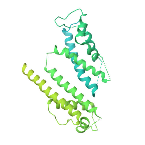
    The hERG channel is a voltage-gated potassium channel involved in cardiac repolarization. Off-target hERG inhibition by drugs has become a critical issue in the pharmaceutical industry. The three-dimensional structure of the hERG channel was recently reported at 3.8-Å resolution using cryogenic electron microscopy (cryo-EM). However, the drug inhibition mechanism remains unclear because of the scarce structural information regarding the drug- and potassium-bound hERG channels. In this study, we obtained the cryo-EM density map of potassium-bound hERG channel complexed with astemizole, a well-known hERG inhibitor that increases risk of potentially fatal arrhythmia, at 3.5-Å resolution. The structure suggested that astemizole inhibits potassium conduction by binding directly below the selectivity filter. Furthermore, we propose a possible binding model of astemizole to the hERG channel and provide insights into the unusual sensitivity of hERG to several drugs.


  • Organizational Affiliation

    Department of Chemistry, Graduate School of Science, Chiba University, 1-33 Yayoi-cho, Inage, Chiba 263-8522, Japan.


Macromolecules
Find similar proteins by:  (by identity cutoff)  |  3D Structure
Entity ID: 1
MoleculeChains Sequence LengthOrganismDetailsImage
potassium channel
A, B, C, D
820Homo sapiensMutation(s): 0 
Entity Groups  
Sequence Clusters30% Identity50% Identity70% Identity90% Identity95% Identity100% Identity
Sequence Annotations
Expand
  • Reference Sequence
Experimental Data & Validation

Experimental Data

  • Method: ELECTRON MICROSCOPY
  • Resolution: 3.70 Å
  • Aggregation State: PARTICLE 
  • Reconstruction Method: SINGLE PARTICLE 
EM Software:
TaskSoftware PackageVersion
MODEL REFINEMENTPHENIX1.16
RECONSTRUCTIONRELION3.0

Structure Validation

View Full Validation Report


Entry History & Funding Information

Deposition Data


Funding OrganizationLocationGrant Number
Japan Agency for Medical Research and Development (AMED)JapanJP20am0101071
Japan Agency for Medical Research and Development (AMED)JapanJP20am0101083
Japan Society for the Promotion of Science (JSPS)Japan18H05425

Revision History  (Full details and data files)

  • Version 1.0: 2021-01-20
    Type: Initial release
  • Version 1.1: 2021-03-17
    Changes: Database references
  • Version 1.2: 2024-03-27
    Changes: Data collection, Database references