7KPP

Structure of the E102A mutant of a GNAT superfamily PA3944 acetyltransferase


Experimental Data Snapshot

  • Method: X-RAY DIFFRACTION
  • Resolution: 1.45 Å
  • R-Value Free: 0.168 
  • R-Value Work: 0.149 
  • R-Value Observed: 0.150 

wwPDB Validation   3D Report Full Report


Ligand Structure Quality Assessment 


This is version 1.2 of the entry. See complete history


Literature

Gcn5-Related N- Acetyltransferases (GNATs) With a Catalytic Serine Residue Can Play Ping-Pong Too.

Baumgartner, J.T.Habeeb Mohammad, T.S.Czub, M.P.Majorek, K.A.Arolli, X.Variot, C.Anonick, M.Minor, W.Ballicora, M.A.Becker, D.P.Kuhn, M.L.

(2021) Front Mol Biosci 8: 646046-646046

  • DOI: https://doi.org/10.3389/fmolb.2021.646046
  • Primary Citation of Related Structures:  
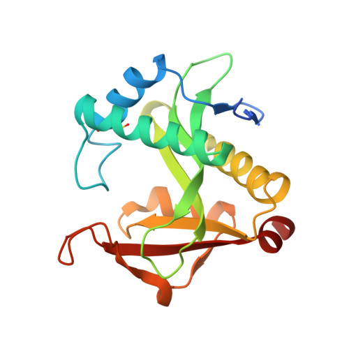
    7KPP, 7KPS

  • PubMed Abstract: 

    Enzymes in the Gcn5-related N- acetyltransferase (GNAT) superfamily are widespread and critically involved in multiple cellular processes ranging from antibiotic resistance to histone modification. While acetyl transfer is the most widely catalyzed reaction, recent studies have revealed that these enzymes are also capable of performing succinylation, condensation, decarboxylation, and methylcarbamoylation reactions. The canonical chemical mechanism attributed to GNATs is a general acid/base mechanism; however, mounting evidence has cast doubt on the applicability of this mechanism to all GNATs. This study shows that the Pseudomonas aeruginosa PA3944 enzyme uses a nucleophilic serine residue and a hybrid ping-pong mechanism for catalysis instead of a general acid/base mechanism. To simplify this enzyme's kinetic characterization, we synthesized a polymyxin B substrate analog and performed molecular docking experiments. We performed site-directed mutagenesis of key active site residues (S148 and E102) and determined the structure of the E102A mutant. We found that the serine residue is essential for catalysis toward the synthetic substrate analog and polymyxin B, but the glutamate residue is more likely important for substrate recognition or stabilization. Our results challenge the current paradigm of GNAT mechanisms and show that this common enzyme scaffold utilizes different active site residues to accomplish a diversity of catalytic reactions.


  • Organizational Affiliation

    Department of Chemistry and Biochemistry, San Francisco State University, San Francisco, CA, United States.


Macromolecules
Find similar proteins by:  (by identity cutoff)  |  3D Structure
Entity ID: 1
MoleculeChains Sequence LengthOrganismDetailsImage
Acetyltransferase PA3944
A, B
194Pseudomonas aeruginosaMutation(s): 1 
Gene Names: PA3944
EC: 2.3.1
UniProt
Find proteins for Q9HX72 (Pseudomonas aeruginosa (strain ATCC 15692 / DSM 22644 / CIP 104116 / JCM 14847 / LMG 12228 / 1C / PRS 101 / PAO1))
Explore Q9HX72 
Go to UniProtKB:  Q9HX72
Entity Groups  
Sequence Clusters30% Identity50% Identity70% Identity90% Identity95% Identity100% Identity
UniProt GroupQ9HX72
Sequence Annotations
Expand
  • Reference Sequence
Small Molecules
Ligands 3 Unique
IDChains Name / Formula / InChI Key2D Diagram3D Interactions
COA (Subject of Investigation/LOI)
Query on COA

Download Ideal Coordinates CCD File 
C [auth A],
O [auth B]
COENZYME A
C21 H36 N7 O16 P3 S
RGJOEKWQDUBAIZ-IBOSZNHHSA-N
EDO
Query on EDO

Download Ideal Coordinates CCD File 
D [auth A]
G [auth A]
H [auth A]
I [auth A]
J [auth A]
D [auth A],
G [auth A],
H [auth A],
I [auth A],
J [auth A],
R [auth B],
S [auth B]
1,2-ETHANEDIOL
C2 H6 O2
LYCAIKOWRPUZTN-UHFFFAOYSA-N
UNX
Query on UNX

Download Ideal Coordinates CCD File 
E [auth A]
F [auth A]
K [auth A]
L [auth A]
M [auth A]
E [auth A],
F [auth A],
K [auth A],
L [auth A],
M [auth A],
N [auth A],
P [auth B],
Q [auth B]
UNKNOWN ATOM OR ION
X
Modified Residues  1 Unique
IDChains TypeFormula2D DiagramParent
CSX
Query on CSX
A, B
L-PEPTIDE LINKINGC3 H7 N O3 SCYS
Experimental Data & Validation

Experimental Data

  • Method: X-RAY DIFFRACTION
  • Resolution: 1.45 Å
  • R-Value Free: 0.168 
  • R-Value Work: 0.149 
  • R-Value Observed: 0.150 
  • Space Group: P 1
  • Diffraction Data: https://doi.org/10.18430/m37kpp
Unit Cell:
Length ( Å )Angle ( ˚ )
a = 36.344α = 98.03
b = 44.038β = 106.88
c = 60.184γ = 90.03
Software Package:
Software NamePurpose
HKL-3000data scaling
REFMACrefinement
PDB_EXTRACTdata extraction
HKL-3000data reduction
MOLREPphasing
Cootmodel building

Structure Validation

View Full Validation Report


Ligand Structure Quality Assessment 


Entry History & Funding Information

Deposition Data


Funding OrganizationLocationGrant Number
National Institutes of Health/National Institute Of Allergy and Infectious Diseases (NIH/NIAID)United States--

Revision History  (Full details and data files)

  • Version 1.0: 2020-11-25
    Type: Initial release
  • Version 1.1: 2023-08-09
    Changes: Database references, Refinement description
  • Version 1.2: 2023-10-18
    Changes: Data collection, Refinement description