7NG5

P1c-state of wild type human mitochondrial LONP1 protease with bound substrate protein in presence of ATP/ADP mix


Experimental Data Snapshot

  • Method: ELECTRON MICROSCOPY
  • Resolution: 3.80 Å
  • Aggregation State: PARTICLE 
  • Reconstruction Method: SINGLE PARTICLE 

wwPDB Validation   3D Report Full Report


This is version 3.2 of the entry. See complete history


Literature

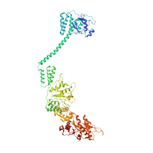
Catalytic cycling of human mitochondrial Lon protease.

Mohammed, I.Schmitz, K.A.Schenck, N.Balasopoulos, D.Topitsch, A.Maier, T.Abrahams, J.P.

(2022) Structure 30: 1254-1268.e7

  • DOI: https://doi.org/10.1016/j.str.2022.06.006
  • Primary Citation of Related Structures:  
    7NFY, 7NG4, 7NG5, 7NGC, 7NGF, 7NGL, 7NGP, 7NGQ, 7OXO

  • PubMed Abstract: 

    The mitochondrial Lon protease (LonP1) regulates mitochondrial health by removing redundant proteins from the mitochondrial matrix. We determined LonP1 in eight nucleotide-dependent conformational states by cryoelectron microscopy (cryo-EM). The flexible assembly of N-terminal domains had 3-fold symmetry, and its orientation depended on the conformational state. We show that a conserved structural motif around T803 with a high similarity to the trypsin catalytic triad is essential for proteolysis. We show that LonP1 is not regulated by redox potential, despite the presence of two conserved cysteines at disulfide-bonding distance in its unfoldase core. Our data indicate how sequential ATP hydrolysis controls substrate protein translocation in a 6-fold binding change mechanism. Substrate protein translocation, rather than ATP hydrolysis, is a rate-limiting step, suggesting that LonP1 is a Brownian ratchet with ATP hydrolysis preventing translocation reversal. 3-fold rocking motions of the flexible N-domain assembly may assist thermal unfolding of the substrate protein.


  • Organizational Affiliation

    Biozentrum, University of Basel, Basel, Switzerland.


Macromolecules
Find similar proteins by:  (by identity cutoff)  |  3D Structure
Entity ID: 1
MoleculeChains Sequence LengthOrganismDetailsImage
Lon protease homolog, mitochondrial
A, B, C, D, E
A, B, C, D, E, F
853Homo sapiensMutation(s): 0 
Gene Names: LONP1PRSS15
EC: 3.4.21.53
UniProt & NIH Common Fund Data Resources
Find proteins for P36776 (Homo sapiens)
Explore P36776 
Go to UniProtKB:  P36776
PHAROS:  P36776
GTEx:  ENSG00000196365 
Entity Groups  
Sequence Clusters30% Identity50% Identity70% Identity90% Identity95% Identity100% Identity
UniProt GroupP36776
Sequence Annotations
Expand
  • Reference Sequence
Find similar proteins by:  (by identity cutoff)  |  3D Structure
Entity ID: 2
MoleculeChains Sequence LengthOrganismDetailsImage
Substrate protein chain:G55Homo sapiensMutation(s): 0 
Sequence Annotations
Expand
  • Reference Sequence
Small Molecules
Ligands 3 Unique
IDChains Name / Formula / InChI Key2D Diagram3D Interactions
ATP (Subject of Investigation/LOI)
Query on ATP

Download Ideal Coordinates CCD File 
H [auth A],
J [auth B],
L [auth C],
O [auth E],
Q [auth F]
ADENOSINE-5'-TRIPHOSPHATE
C10 H16 N5 O13 P3
ZKHQWZAMYRWXGA-KQYNXXCUSA-N
ADP (Subject of Investigation/LOI)
Query on ADP

Download Ideal Coordinates CCD File 
N [auth D]ADENOSINE-5'-DIPHOSPHATE
C10 H15 N5 O10 P2
XTWYTFMLZFPYCI-KQYNXXCUSA-N
MG (Subject of Investigation/LOI)
Query on MG

Download Ideal Coordinates CCD File 
I [auth A],
K [auth B],
M [auth C],
P [auth E],
R [auth F]
MAGNESIUM ION
Mg
JLVVSXFLKOJNIY-UHFFFAOYSA-N
Experimental Data & Validation

Experimental Data

  • Method: ELECTRON MICROSCOPY
  • Resolution: 3.80 Å
  • Aggregation State: PARTICLE 
  • Reconstruction Method: SINGLE PARTICLE 
EM Software:
TaskSoftware PackageVersion
RECONSTRUCTIONRELION
RECONSTRUCTIONcryoSPARC

Structure Validation

View Full Validation Report


Entry History 

Deposition Data

Revision History  (Full details and data files)

  • Version 1.0: 2021-02-24
    Type: Initial release
  • Version 1.1: 2021-04-28
    Changes: Database references
  • Version 2.0: 2021-06-30
    Type: Coordinate replacement
    Reason: Model completeness
    Changes: Advisory, Atomic model, Data collection, Database references, Derived calculations, Structure summary
  • Version 3.0: 2022-08-10
    Changes: Atomic model, Database references
  • Version 3.1: 2022-09-14
    Changes: Database references
  • Version 3.2: 2022-11-09
    Changes: Database references