6T7L

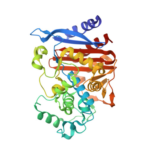
Crystal structure of AmpC from E.coli with Nacubactam (OP0595)


Experimental Data Snapshot

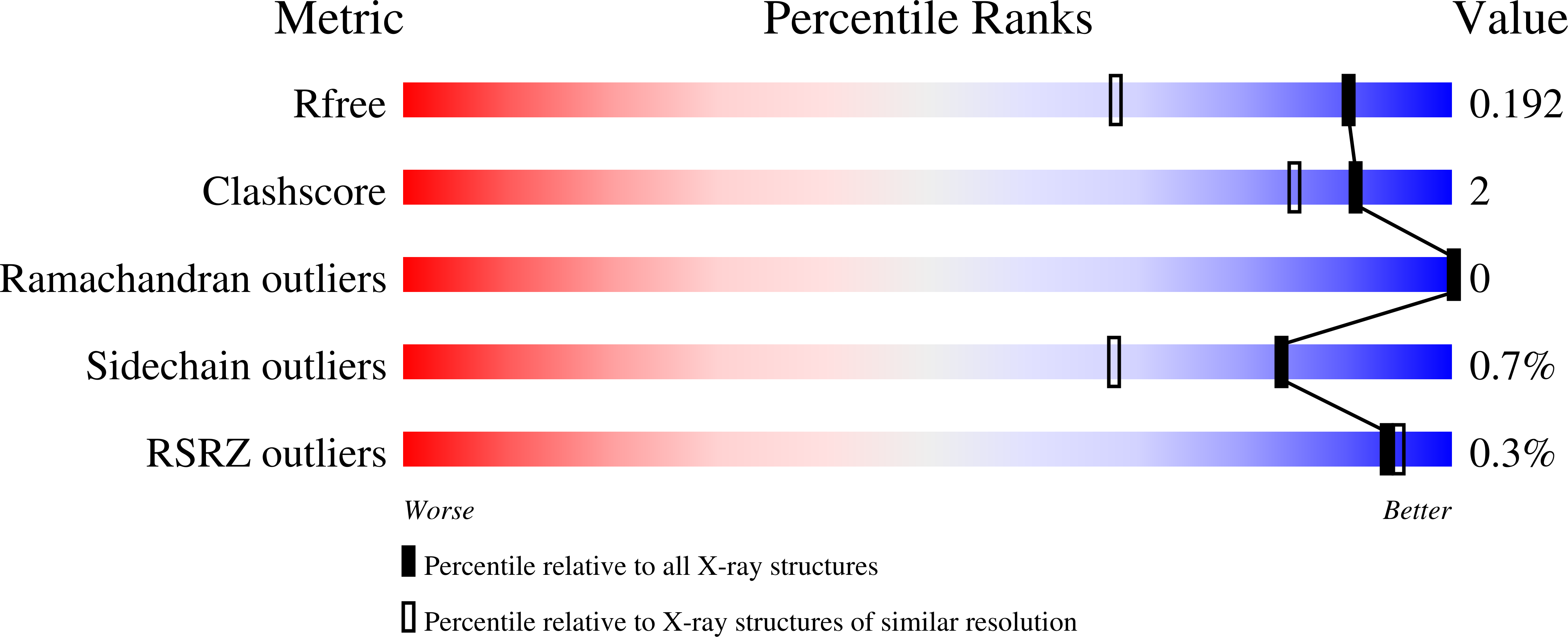
  • Method: X-RAY DIFFRACTION
  • Resolution: 1.47 Å
  • R-Value Free: 0.182 
  • R-Value Work: 0.155 
  • R-Value Observed: 0.156 

Starting Model: experimental
View more details

wwPDB Validation   3D Report Full Report


Ligand Structure Quality Assessment 


This is version 1.4 of the entry. See complete history


Literature

Structural Investigations of the Inhibition of Escherichia coli AmpC beta-Lactamase by Diazabicyclooctanes.

Lang, P.A.Leissing, T.M.Page, M.G.P.Schofield, C.J.Brem, J.

(2021) Antimicrob Agents Chemother 65

  • DOI: https://doi.org/10.1128/AAC.02073-20
  • Primary Citation of Related Structures:  
    6T5Y, 6T7L, 6TBW, 6TPM

  • PubMed Abstract: 

    β-Lactam antibiotics are presently the most important treatments for infections by pathogenic Escherichia coli , but their use is increasingly compromised by β-lactamases, including the chromosomally encoded class C AmpC serine-β-lactamases (SBLs). The diazabicyclooctane (DBO) avibactam is a potent AmpC inhibitor; the clinical success of avibactam combined with ceftazidime has stimulated efforts to optimize the DBO core. We report kinetic and structural studies, including four high-resolution crystal structures, concerning inhibition of the AmpC serine-β-lactamase from E. coli (AmpC EC ) by clinically relevant DBO-based inhibitors: avibactam, relebactam, nacubactam, and zidebactam. Kinetic analyses and mass spectrometry-based assays were used to study their mechanisms of AmpC EC inhibition. The results reveal that, under our assay conditions, zidebactam manifests increased potency (apparent inhibition constant [ K iapp ], 0.69 μM) against AmpC EC compared to that of the other DBOs ( K iapp = 5.0 to 7.4 μM) due to an ∼10-fold accelerated carbamoylation rate. However, zidebactam also has an accelerated off-rate, and with sufficient preincubation time, all the DBOs manifest similar potencies. Crystallographic analyses indicate a greater conformational freedom of the AmpC EC -zidebactam carbamoyl complex compared to those for the other DBOs. The results suggest the carbamoyl complex lifetime should be a consideration in development of DBO-based SBL inhibitors for the clinically important class C SBLs.


  • Organizational Affiliation

    Department of Chemistry, Chemistry Research Laboratory, University of Oxford, Oxford, United Kingdom.


Macromolecules
Find similar proteins by:  (by identity cutoff)  |  3D Structure
Entity ID: 1
MoleculeChains Sequence LengthOrganismDetailsImage
Beta-lactamase358Escherichia coli K-12Mutation(s): 0 
Gene Names: ampCampAb4150JW4111
EC: 3.5.2.6
UniProt
Find proteins for P00811 (Escherichia coli (strain K12))
Explore P00811 
Go to UniProtKB:  P00811
Entity Groups  
Sequence Clusters30% Identity50% Identity70% Identity90% Identity95% Identity100% Identity
UniProt GroupP00811
Sequence Annotations
Expand
  • Reference Sequence
Small Molecules
Ligands 6 Unique
IDChains Name / Formula / InChI Key2D Diagram3D Interactions
OP0 (Subject of Investigation/LOI)
Query on OP0

Download Ideal Coordinates CCD File 
C [auth A](2S,5R)-N-(2-aminoethoxy)-1-formyl-5-[(sulfooxy)amino]piperidine-2-carboxamide
C9 H18 N4 O7 S
YTAWXXHGLPATIN-SFYZADRCSA-N
MES
Query on MES

Download Ideal Coordinates CCD File 
B [auth A]2-(N-MORPHOLINO)-ETHANESULFONIC ACID
C6 H13 N O4 S
SXGZJKUKBWWHRA-UHFFFAOYSA-N
PGE
Query on PGE

Download Ideal Coordinates CCD File 
G [auth A]TRIETHYLENE GLYCOL
C6 H14 O4
ZIBGPFATKBEMQZ-UHFFFAOYSA-N
SO4
Query on SO4

Download Ideal Coordinates CCD File 
F [auth A]SULFATE ION
O4 S
QAOWNCQODCNURD-UHFFFAOYSA-L
ZN
Query on ZN

Download Ideal Coordinates CCD File 
D [auth A],
E [auth A]
ZINC ION
Zn
PTFCDOFLOPIGGS-UHFFFAOYSA-N
CL
Query on CL

Download Ideal Coordinates CCD File 
H [auth A]CHLORIDE ION
Cl
VEXZGXHMUGYJMC-UHFFFAOYSA-M
Experimental Data & Validation

Experimental Data

  • Method: X-RAY DIFFRACTION
  • Resolution: 1.47 Å
  • R-Value Free: 0.182 
  • R-Value Work: 0.155 
  • R-Value Observed: 0.156 
  • Space Group: P 43 3 2
Unit Cell:
Length ( Å )Angle ( ˚ )
a = 138.178α = 90
b = 138.178β = 90
c = 138.178γ = 90
Software Package:
Software NamePurpose
XDSdata reduction
Aimlessdata scaling
PHASERphasing
PHENIXrefinement
PDB_EXTRACTdata extraction

Structure Validation

View Full Validation Report


Ligand Structure Quality Assessment 


Entry History & Funding Information

Deposition Data


Funding OrganizationLocationGrant Number
Medical Research Council (United Kingdom)United KingdomMRF-145-0004-TPG-AVISO

Revision History  (Full details and data files)

  • Version 1.0: 2020-11-18
    Type: Initial release
  • Version 1.1: 2020-12-02
    Changes: Database references
  • Version 1.2: 2021-02-03
    Changes: Database references
  • Version 1.3: 2024-01-24
    Changes: Data collection, Database references, Refinement description
  • Version 1.4: 2024-11-20
    Changes: Structure summary