7UQW

PCC6803 Cyanophycinase S132DAP covalently bound to cyanophycin dimer


Experimental Data Snapshot

  • Method: X-RAY DIFFRACTION
  • Resolution: 1.50 Å
  • R-Value Free: 0.199 
  • R-Value Work: 0.181 
  • R-Value Observed: 0.182 

wwPDB Validation   3D Report Full Report


Ligand Structure Quality Assessment 


This is version 1.1 of the entry. See complete history


Literature

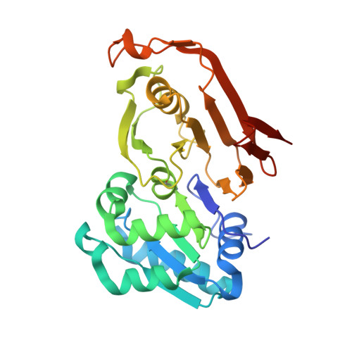
The structure of cyanophycinase in complex with a cyanophycin degradation intermediate.

Sharon, I.Grogg, M.Hilvert, D.Schmeing, T.M.

(2022) Biochim Biophys Acta Gen Subj 1866: 130217-130217

  • DOI: https://doi.org/10.1016/j.bbagen.2022.130217
  • Primary Citation of Related Structures:  
    7UQV, 7UQW

  • PubMed Abstract: 

    Cyanophycinases are serine protease family enzymes which are required for the metabolism of cyanophycin, the natural polymer multi-L-arginyl-poly(L-aspartic acid). Cyanophycinases degrade cyanophycin to β-Asp-Arg dipeptides, which enables use of this important store of fixed nitrogen.


  • Organizational Affiliation

    Department of Biochemistry and Centre de recherche en biologie structurale, McGill University, Montréal, QC H3G 0B1, Canada.


Macromolecules
Find similar proteins by:  (by identity cutoff)  |  3D Structure
Entity ID: 1
MoleculeChains Sequence LengthOrganismDetailsImage
Cyanophycinase
A, B, C
276Synechocystis sp. PCC 6803Mutation(s): 1 
Gene Names: cphBslr2001
EC: 3.4.15.6
UniProt
Find proteins for P73832 (Synechocystis sp. (strain PCC 6803 / Kazusa))
Explore P73832 
Go to UniProtKB:  P73832
Entity Groups  
Sequence Clusters30% Identity50% Identity70% Identity90% Identity95% Identity100% Identity
UniProt GroupP73832
Sequence Annotations
Expand
  • Reference Sequence
Small Molecules
Ligands 3 Unique
IDChains Name / Formula / InChI Key2D Diagram3D Interactions
7ID (Subject of Investigation/LOI)
Query on 7ID

Download Ideal Coordinates CCD File 
D [auth A]
E [auth A]
G [auth B]
H [auth B]
N [auth C]
D [auth A],
E [auth A],
G [auth B],
H [auth B],
N [auth C],
O [auth C]
(2~{S})-4-[[(2~{S})-5-[[azanyl($l^{4}-azanylidene)methyl]amino]-1-$l^{1}-oxidanyl-1-oxidanylidene-pentan-2-yl]amino]-2-$l^{2}-azanyl-4-oxidanylidene-butanoic acid
C10 H19 N5 O5
QCGCETFHYOEVAI-WDSKDSINSA-N
SO4
Query on SO4

Download Ideal Coordinates CCD File 
F [auth A]SULFATE ION
O4 S
QAOWNCQODCNURD-UHFFFAOYSA-L
ARF
Query on ARF

Download Ideal Coordinates CCD File 
I [auth B]
J [auth B]
K [auth B]
L [auth B]
M [auth B]
I [auth B],
J [auth B],
K [auth B],
L [auth B],
M [auth B],
P [auth C],
Q [auth C]
FORMAMIDE
C H3 N O
ZHNUHDYFZUAESO-UHFFFAOYSA-N
Modified Residues  1 Unique
IDChains TypeFormula2D DiagramParent
DPP
Query on DPP
A, B, C
L-PEPTIDE LINKINGC3 H8 N2 O2ALA
Experimental Data & Validation

Experimental Data

  • Method: X-RAY DIFFRACTION
  • Resolution: 1.50 Å
  • R-Value Free: 0.199 
  • R-Value Work: 0.181 
  • R-Value Observed: 0.182 
  • Space Group: C 2 2 21
Unit Cell:
Length ( Å )Angle ( ˚ )
a = 76.463α = 90
b = 132.947β = 90
c = 164.092γ = 90
Software Package:
Software NamePurpose
REFMACrefinement
PHENIXrefinement
XDSdata reduction
pointlessdata scaling
PHASERphasing

Structure Validation

View Full Validation Report


Ligand Structure Quality Assessment 


Entry History & Funding Information

Deposition Data


Funding OrganizationLocationGrant Number
Canadian Institutes of Health Research (CIHR)Canada178084

Revision History  (Full details and data files)

  • Version 1.0: 2022-08-10
    Type: Initial release
  • Version 1.1: 2023-10-18
    Changes: Data collection, Refinement description