Structure Determination MethodologyScientific Name of Source OrganismMore... Refinement Resolution (Å)Enzyme Classification Name | Bagu, J.R., Sykes, B.D. (1995) Nat Struct Biol 2: 114-116 | Released | 1996-11-08 | | Method | SOLUTION NMR | | Organisms | | | Macromolecule | |
Bagu, J.R., Sykes, B.D. (1995) Nat Struct Biol 2: 114-116 | Released | 1996-11-08 | | Method | SOLUTION NMR | | Organisms | | | Macromolecule | |
Bagu, J.R., Sykes, B.D. (1995) Nat Struct Biol 2: 114-116 | Released | 1996-11-08 | | Method | SOLUTION NMR | | Organisms | | | Macromolecule | |
Bagu, J.R., Sykes, B.D. (1995) Nat Struct Biol 2: 114-116 | Released | 1996-11-08 | | Method | SOLUTION NMR | | Organisms | | | Macromolecule | |
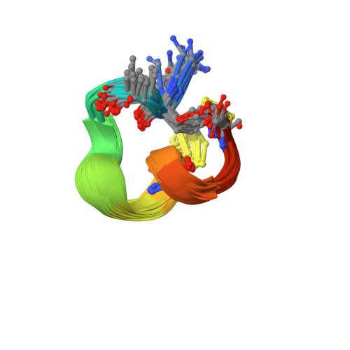
Trogen, G., Zdunek, J. (1996) Biochemistry 35: 3197-3205 | Released | 1996-12-07 | | Method | SOLUTION NMR | | Organisms | | | Macromolecule | |
Maynes, J.T., Luu, H.A., Cherney, M.M., Andersen, R.J., Williams, D., Holmes, C.F., James, M.N. (2006) J Mol Biology 356: 111-120 | Released | 2006-01-17 | | Method | X-RAY DIFFRACTION 2.1 Å | | Organisms | | | Macromolecule | | | Unique Ligands | BME, MN |
Maynes, J.T., Luu, H.A., Cherney, M.M., Andersen, R.J., Williams, D., Holmes, C.F., James, M.N. (2006) J Mol Biology 356: 111-120 | Released | 2006-01-17 | | Method | X-RAY DIFFRACTION 2.3 Å | | Organisms | | | Macromolecule | | | Unique Ligands | MN |
Cho, U.S., Xu, W. (2007) Nature 445: 53-57 | Released | 2006-12-26 | | Method | X-RAY DIFFRACTION 3.5 Å | | Organisms | | | Macromolecule | | | Unique Ligands | MN |
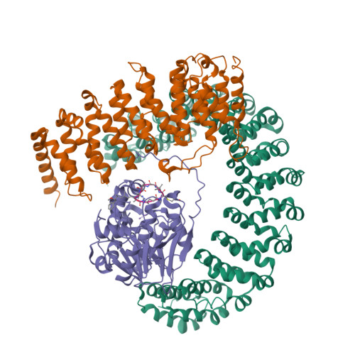
Xing, Y., Xu, Y., Chen, Y., Jeffrey, P.D., Chao, Y., Shi, Y. (2006) Cell 127: 341-353 | Released | 2006-11-07 | | Method | X-RAY DIFFRACTION 2.8 Å | | Organisms | | | Macromolecule | | | Unique Ligands | MN |
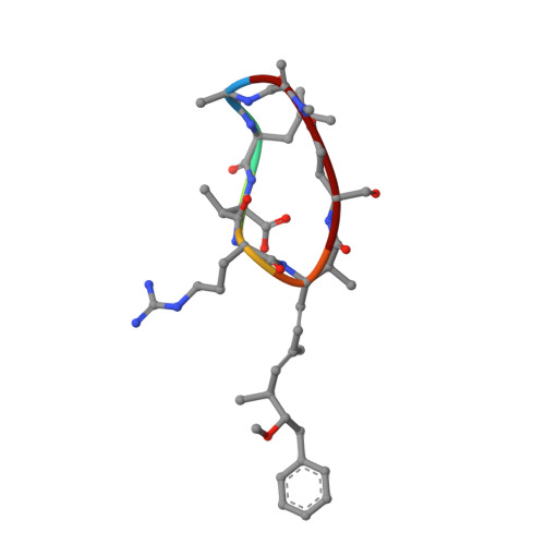
Heinis, C., Chen, S. (2014) Nat Chem 6: 1009-1016 | Released | 2014-09-24 | | Method | SOLUTION NMR | | Organisms | | | Macromolecule | |
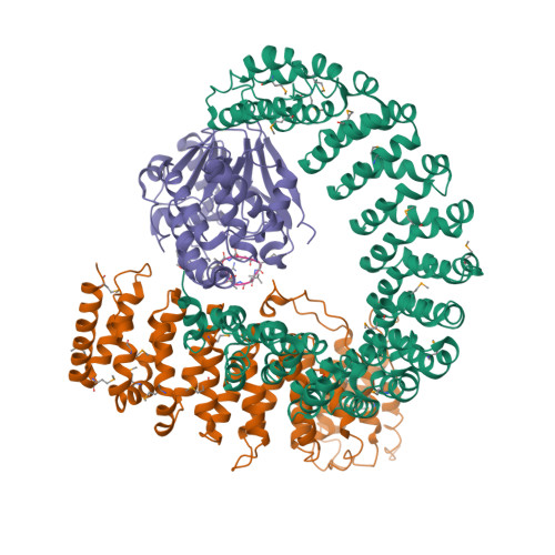
Xing, Y., Xu, Y., Chen, Y., Chao, Y., Lin, Z., Shi, Y. (2006) Cell 127: 1239-1251 | Released | 2006-12-12 | | Method | X-RAY DIFFRACTION 3.8 Å | | Organisms | | | Macromolecule | | | Unique Ligands | MN |
Chen, Y., Xing, Y., Xu, Y., Chao, Y., Lin, Z., Jeffrey, P.D., Shi, Y. (2006) Cell 127: 1239-1251 | Released | 2006-12-12 | | Method | X-RAY DIFFRACTION 3.6 Å | | Organisms | | | Macromolecule | | | Unique Ligands | MN |
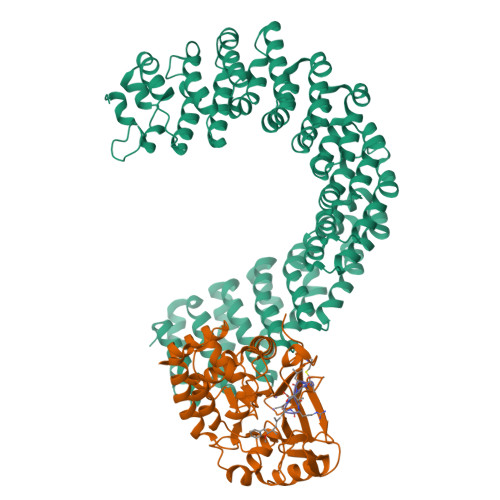
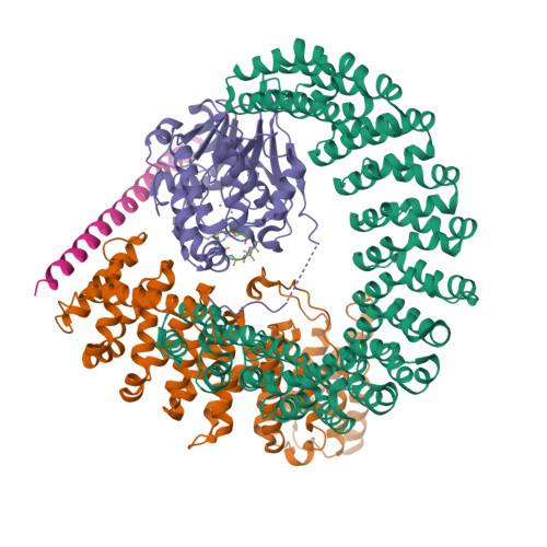
Xu, Z., Xu, W. (2009) Mol Cell 35: 426-441 | Released | 2009-09-22 | | Method | X-RAY DIFFRACTION 2.7 Å | | Organisms | | | Macromolecule | | | Unique Ligands | MN |
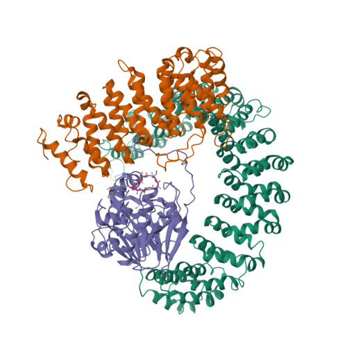
Wlodarchak, N., Satyshur, K.A., Guo, F., Xing, Y. (2013) Cell Res 23: 931-946 | Released | 2013-05-08 | | Method | X-RAY DIFFRACTION 2.43 Å | | Organisms | | | Macromolecule | | | Unique Ligands | CA, MLI, MN, PEG |
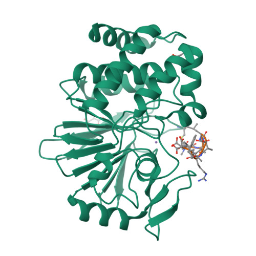
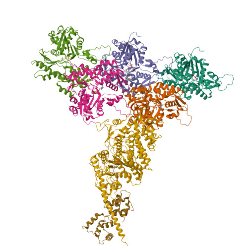
Mentes, A., Huehn, A., Liu, X., Zwolak, A., Dominguez, R., Shuman, H., Ostap, E.M., Sindelar, C.V. (2018) Proc Natl Acad Sci U S A 115: 1292-1297 | Released | 2018-01-31 | | Method | ELECTRON MICROSCOPY 3.2 Å | | Organisms | | | Macromolecule | | | Unique Ligands | ADP, MG |
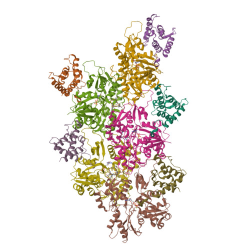
Iwamoto, D.V., Huehn, A.R., Simon, B., Huet-Calderwood, C., Baldassarre, M., Sindelar, C.V., Calderwood, D.A. (2018) Nat Struct Mol Biol 25: 918-927 | Released | 2018-09-19 | | Method | ELECTRON MICROSCOPY 3.54 Å | | Organisms | | | Macromolecule | | | Unique Ligands | ADP, MG |
|