Structure Determination MethodologyScientific Name of Source OrganismMore... Refinement Resolution (Å)Enzyme Classification NameMembrane Protein Annotation | Asojo, O.A., Gulnik, S.V., Afonina, E., Yu, B., Ellman, J.A., Haque, T.S., Silva, A.M. (2003) J Mol Biology 327: 173-181 | Released | 2002-06-12 | | Method | X-RAY DIFFRACTION 2.8 Å | | Organisms | | | Macromolecule | |
Lindberg, J., Johansson, P.-O., Rosenquist, A., Kvarnstroem, I., Vrang, L., Samuelsson, B., Unge, T. To be published | Released | 2006-07-05 | | Method | X-RAY DIFFRACTION 2.7 Å | | Organisms | | | Macromolecule | |
Prade, L. To be published | Released | 2005-08-23 | | Method | X-RAY DIFFRACTION 1.7 Å | | Organisms | | | Macromolecule | |
Yang, J., Quail, J.W. (1999) Acta Crystallogr D Biol Crystallogr 55: 625-630 | Released | 1997-09-17 | | Method | X-RAY DIFFRACTION 2.7 Å | | Organisms | | | Macromolecule | | | Unique Ligands | NAG | | Unique branched monosaccharides | BMA, NAG |
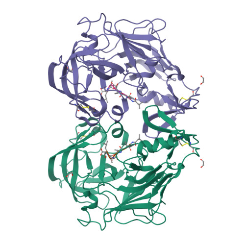
Nascimento, A.S., Krauchenco, S., Golubev, A.M., Gustchina, A., Wlodawer, A., Polikarpov, I. (2008) J Mol Biology 382: 763-778 | Released | 2008-10-07 | | Method | X-RAY DIFFRACTION 1.85 Å | | Organisms | | | Macromolecule | |
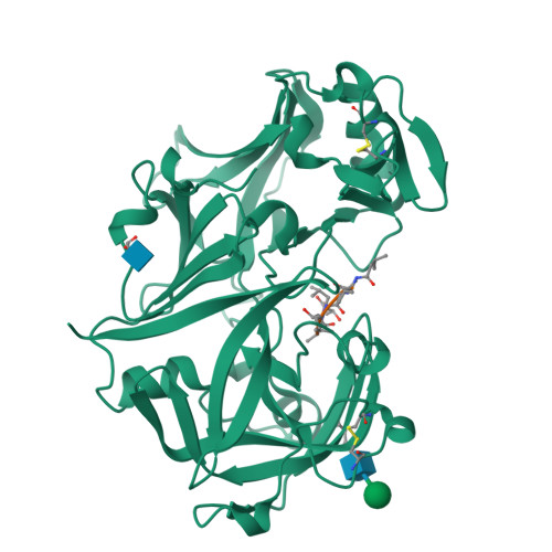
Dostal, J., Brynda, J., Hruskova-Heidingsfeldova, O., Sieglova, I., Pichova, I., Rezacova, P. (2009) J Struct Biol 167: 145-152 | Released | 2009-05-19 | | Method | X-RAY DIFFRACTION 1.85 Å | | Organisms | | | Macromolecule | | | Unique Ligands | GOL, SO4 |
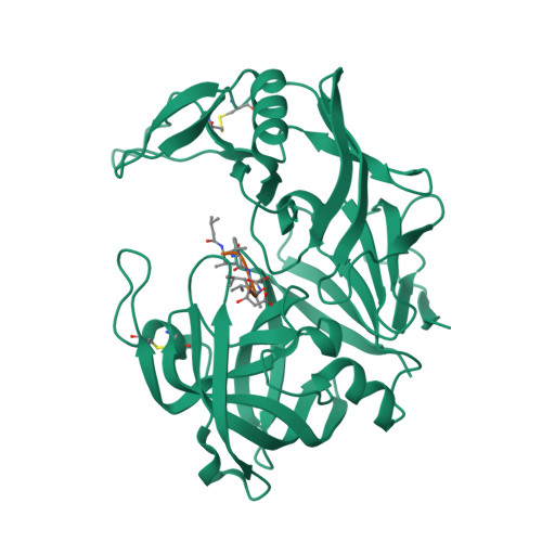
Tiefenbrunn, T., Stout, C.D. (2013) ACS Chem Biol 8: 1223-1231 | Released | 2013-05-01 | | Method | X-RAY DIFFRACTION 1.794 Å | | Organisms | | | Macromolecule | | | Unique Ligands | IOP |
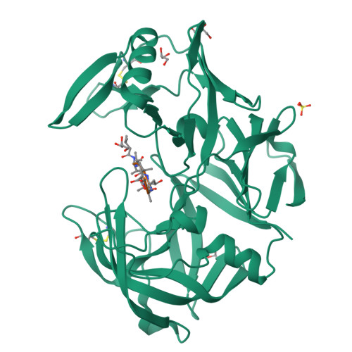
Dostal, J., Brynda, J., Hruskova-Heidingsfeldova, O., Rezacova, P., Mareckova, L., Pichova, I. (2015) Acta Crystallogr D Biol Crystallogr 71: 2494-2504 | Released | 2016-11-30 | | Method | X-RAY DIFFRACTION 0.83 Å | | Organisms | | | Macromolecule | |
Asojo, O.A. To be published | Released | 2016-03-09 | | Method | X-RAY DIFFRACTION 3 Å | | Organisms | | | Macromolecule | |
Wu, Y., Batyuk, A., Mittl, P.R., Honegger, A., Plueckthun, A. (2018) J Mol Biology 430: 2128-2138 | Released | 2017-12-13 | | Method | X-RAY DIFFRACTION 3.2 Å | | Organisms | | | Macromolecule | | | Unique Ligands | ADN, SO4 |
|