


Full Text |
QUERY: BIRD Name HAS EXACT PHRASE "=alpha-cyclodextrin" | MyPDB Login | Search API |
| Search Summary | This query matches 22 Structures. |
Structure Determination MethodologyScientific Name of Source OrganismMore... TaxonomyExperimental MethodPolymer Entity TypeRefinement Resolution (Å)Release DateEnzyme Classification NameMembrane Protein AnnotationSymmetry TypeSCOP Classification | 1 to 22 of 22 Structures Page 1 of 1 Sort by 
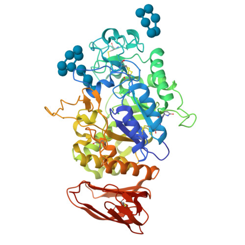
THREE-DIMENSIONAL STRUCTURE OF SOYBEAN BETA-AMYLASE DETERMINED AT 3.0 ANGSTROMS RESOLUTION: PRELIMINARY CHAIN TRACING OF THE COMPLEX WITH ALPHA-CYCLODEXTRINMikami, B., Hehre, E.J., Sato, M., Katsube, Y., Hirose, M., Morita, Y., Sacchettini, J.C. (1993) Biochemistry 32: 6836-6845

COMPLEX OF A (D229N/E257Q) DOUBLE MUTANT CGTASE FROM BACILLUS CIRCULANS STRAIN 251 WITH MALTOTETRAOSE AT 120 K AND PH 9.1 OBTAINED AFTER SOAKING THE CRYSTAL WITH ALPHA-CYCLODEXTRINKnegtel, R.M.A., Strokopytov, B.V., Dijkstra, B.W. (1995) J Biological Chem 270: 29256-29264
Crystal structure of Thermoactinomyces vulgaris R-47 alpha-amylase 2/alpha-cyclodextrin complexOhtaki, A., Mizuno, M., Tonozuka, T., Sakano, Y., Kamitori, S. (2004) J Biological Chem 279: 31033-31040

Crystal structure of pullulanase type I from Bacillus subtilis str. 168 complexed with alpha-cyclodextrinMikami, B., Malle, D., Utsumi, S., Iwamoto, H., Katsuya, Y. To be published

Crystal structure of Barley Beta-Amylase complexed with alpha- cyclodextrinRejzek, M., Stevenson, C.E.M., Southard, A.M., Stanley, D., Denyer, K., Smith, A.M., Naldrett, M.J., Lawson, D.M., Field, R.A. (2011) Mol Biosyst 7: 718

BARLEY LIMIT DEXTRINASE IN COMPLEX WITH ALPHA-CYCLODEXTRINVester-Christensen, M.B., Hachem, M.A., Svensson, B., Henriksen, A. (2010) J Mol Biology 403: 739
Crystal structure of cyclo/maltodextrin-binding protein complexed with alpha-cyclodextrinMatsumoto, M., Yamada, M., Kurakata, Y., Yoshida, H., Kamitori, S., Nishikawa, A., Tonozuka, T. (2009) FEBS J 276: 3008-3019

Alpha-amylase B in complex with maltotetraose and alpha-cyclodextrinTan, T.-C., Mijts, B.N., Swaminathan, K., Patel, B.K.C., Divne, C. (2008) J Mol Biology 378: 850-868

B. thetaiotaomicron SusD with alpha-cyclodextrinKoropatkin, N.M., Martens, E.C., Gordon, J.I., Smith, T.J. (2008) Structure 16: 1105-1115
Structural base for cyclodextrin hydrolysisBuedenbender, S., Schulz, G.E. (2009) J Mol Biology 385: 606-617
Structural base for cyclodextrin hydrolysisBuedenbender, S., Schulz, G.E. (2009) J Mol Biology 385: 606-617
X-ray Crystallographic Analysis of Pig Pancreatic Alpha-Amylase with Alpha-cyclodextrinLarson, S.B., Day, J.S., McPherson, A. (2010) Biochemistry 49: 3101-3115

GlgE isoform 1 from Streptomyces coelicolor with alpha-cyclodextrin boundSyson, K., Stevenson, C.E.M., Rejzek, M., Fairhurst, S.A., Nair, A., Bruton, C.J., Field, R.A., Chater, K.F., Lawson, D.M., Bornemann, S. (2011) J Biological Chem 286: 38298

GlgE isoform 1 from Streptomyces coelicolor with alpha-cyclodextrin and maltose boundSyson, K., Stevenson, C.E.M., Rejzek, M., Fairhurst, S.A., Nair, A., Bruton, C.J., Field, R.A., Chater, K.F., Lawson, D.M., Bornemann, S. (2011) J Biological Chem 286: 38298
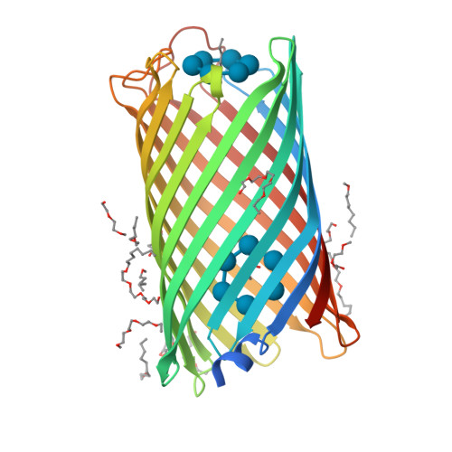
Crystal structure of CymA from Klebsiella oxytocavan den Berg, B., Bhamidimarri, S.P., Kleinekathoefer, U., Winterhalter, M. (2015) Proc Natl Acad Sci U S A 112: E2991-E2999
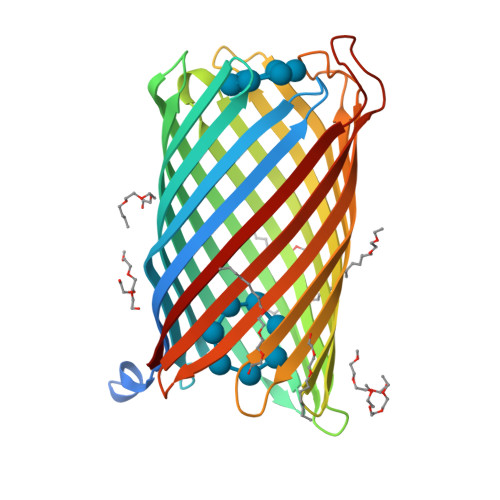
Crystal structure of CymA from Klebsiella oxytocavan den Berg, B., Bhamidimarri, S.P., Kleinekathoefer, U., Winterhalter, M. (2015) Proc Natl Acad Sci U S A 112: E2991-E2999
Structure of SusE with alpha-cyclodextrinKoropatkin, N.M., Cameron, E.A., Martens, E.C. (2012) J Biological Chem 287: 34614-34625
Crystal structure of E.Coli branching enzyme in complex with alpha cyclodextrinFeng, L., Nosrati, M., Geiger, J.H. (2016) Acta Crystallogr D Struct Biol 72: 641-647

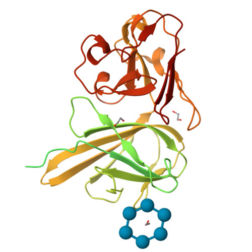
Maltodextrin binding protein MalE1 from L. casei BL23 bound to alpha-cyclodextrinHomburg, C., Bommer, M., Wuttge, S., Hobe, C., Beck, S., Dobbek, H., Deutscher, J., Licht, A., Schneider, E. (2017) Mol Microbiol 105: 25-45
Crystal structure of Pullulanase from Klebsiella pneumoniae complex at 1 mM alpha-cyclodextrinSaka, N., Iwamoto, H., Takahashi, N., Mizutani, K., Mikami, B. (2018) Acta Crystallogr D Struct Biol 74: 1115-1123
Crystal structure of Pullulanase from Klebsiella pneumoniae complex at 10 mM alpha-cyclodextrinSaka, N., Iwamoto, H., Takahashi, N., Mizutani, K., Mikami, B. (2018) Acta Crystallogr D Struct Biol 74: 1115-1123
GH57 family amylopullulanase D352N mutant from Aquifex aeolicus complex with alpha-cyclodextrinZhu, Z.M., Wang, W.W., Li, M.J., Xu, Q., Zhou, H., Huang, L.Q., Wang, Q.S., Yu, F. (2025) J Struct Biol 217: 108199-108199
1 to 22 of 22 Structures Page 1 of 1 Sort by |


