Warning You are using a web browser that we do not support. Our website will not work properly. Please update to a newer version or download a new web browser, such as Chrome or Firefox.
Help
Contact us
  • Sign in with Google
  • Sign in with Facebook
  • Sign in with ORCID

  • Help
RCSB PDB
  • Deposit 

      Prepare Data

    • PDBx/mmCIF file
    • pdb_extract
    • SF-Tool
    • Ligand Expo
    • MAXIT

      Validate Data

    • Validation Server
    • Validation API
    • Information for Journals
    • Validation Task Forces

      Deposit Data

    • wwPDB OneDep System
    • PDB-IHM

      Help and Resources

    • Deposit FAQ
    • Validation FAQ
    • Tutorials
    • Annotation Policies
    • Processing Procedures
    • PDBx/mmCIF Dictionary
    • PDBx/mmCIF User Guide
    • Chemical Component Dictionary
    • Biologically Interesting Molecule Reference Dictionary (BIRD)
    • BioSync/Beamlines/Facilities
    • Related Tools
  • Search 
    • Advanced Search
    • Sequence Similarity Search
    • Structure Similarity Search
    • Chemical Similarity Search
    • Chemical Sketch Tool
    • Browse by Annotations
    • New Entries
    • Unreleased Entries
    • PDB Statistics
  • Visualize 
    • Mol* (MolStar)
    • Sequence Annotations Viewer
    • Genome View
  • Analyze 
    • Pairwise Structure Alignment
    • Symmetry Resources in the PDB
    • Structure Quality
    • Grouping Structures
    • PDB Citation MeSH Network Explorer
    • PDB Statistics
    • EPPIC Biological Assemblies
    • External Data and Resources
    • Integrated Resources
    • Additional Resources
  • Download 
    • Coordinates and Experimental Data
    • Sequences
    • Ligands
    • File Download Services
    • Web APIs
  • Learn 

    • Training Courses
    • Guide to PDB Data
    • Molecule of the Month
    • Educational Resources
    • Curricula
    • Browse
    • News
    • SciArt Galleries
    • Irving Geis
    • David Goodsell
  • About 
    • Contact Us
    • About RCSB PDB
    • Vision and Mission
    • Citation, Usage, Privacy Policies, Logo
    • News
    • PDB History
    • PDB50
    • User Community
    • Publications
    • RCSB PDB Advisory Committee
    • Team Members
    • Service Status
  • Careers
  • COVID-19
RCSB PDB
236,974
Structures from the PDB archive
1,068,577
Computed Structure Models (CSM)
Include CSM
Advanced Search
|
Browse Annotations
Help
  • PDB-101
  • wwPDB
  • EMDataResource
  • NAKB: Nucleic Acid Knowledgebase
  • wwPDB Foundation
  • PDB-IHM Logo
  • Search
  • Query History
  • Browse Annotations
  • MyPDB
QUERY: BIRD Name HAS EXACT PHRASE "=beta-turanose"
MyPDB Login
 Search API
Advanced Search Query Builder
Help
Full Text
Structure Attributes
Chemical Attributes
Help
x
NOT
Count
Add Subquery
Sequence Similarity
Sequence Motif
Structure Similarity
Structure Motif
Chemical Similarity
Return
grouped by
Include Computed Structure Models (CSM)
Count
Clear
Search
Search Summary This query matches 3 Structures.
Refinements
Structure Determination Methodology
Scientific Name of Source Organism
Taxonomy
Experimental Method
Polymer Entity Type
Refinement Resolution (Å)
Release Date
Enzyme Classification Name
Symmetry Type
SCOP Classification
1 to 3 of 3 Structures
Page 1 of 1
Sort by
Gallery item
 Explore in 3D

1N3Q | pdb_00001n3q

Download FileView File

Pterocarpus angolensis lectin complexed with turanose

Loris, R., Imberty, A., Beeckmans, S., Van Driessche, E., Read, J.S., Bouckaert, J., De Greve, H., Buts, L., Wyns, L.

(2003) J Biological Chem 278: 16297-16303

Released2002-11-20
Method
X-RAY DIFFRACTION 2.2 Å
Organisms
Pterocarpus angolensis
Macromolecule
lectin PAL (protein)
Unique LigandsCA, MN
Unique branched monosaccharidesBDF, FRU, GLC
Gallery item
 Explore in 3D

3UEQ | pdb_00003ueq

Download FileView File

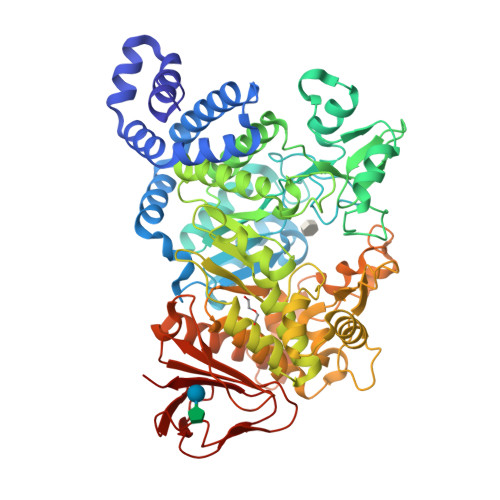
Crystal structure of amylosucrase from Neisseria polysaccharea in complex with turanose

Guerin, F., Pizzut-Serin, S., Potocki-Veronese, G., Guillet, V., Mourey, L., Remaud-Simeon, M., Andre, I., Tranier, S.

(2012) J Biological Chem 287: 6642-6654

Released2012-01-11
Method
X-RAY DIFFRACTION 1.85 Å
Organisms
Neisseria polysaccharea
Macromolecule
Amylosucrase (protein)
Unique LigandsOTU, PEG
Unique branched monosaccharidesFRU, GLC
Gallery item
 Explore in 3D

3UER | pdb_00003uer

Download FileView File

Crystal structure of amylosucrase from Deinococcus geothermalis in complex with turanose

Guerin, F., Pizzut-Serin, S., Potocki-Veronese, G., Guillet, V., Mourey, L., Remaud-Simeon, M., Andre, I., Tranier, S.

(2012) J Biological Chem 287: 6642-6654

Released2012-01-11
Method
X-RAY DIFFRACTION 2.1 Å
Organisms
Deinococcus geothermalis DSM 11300
Macromolecule
Amylosucrase (protein)
Unique branched monosaccharidesFRU, GLC, Z9N
1 to 3 of 3 Structures
Page 1 of 1
Sort by
  • About
  • About Us
  • Citing Us
  • Publications
  • Team
  • Careers
  • Usage & Privacy
  • Support
  • Contact Us
  • Help
  • Website FAQ
  • Glossary
  • Service Status
  • RCSB PDB is hosted by

  • The Rutgers Artificial Intelligence and Data Science Collaboratory logo
    Uiversity of California San Diego logoSan Diego Supercomputer Center logo
    University of California San Francisco Logo
  • RCSB PDB is a member of the

  • wwPDB
    EMDataResource

  • RCSB Partners
  • Nucleic Acid Knowledgebase
  • wwPDB Partners
  • RCSB PDB
  • PDBe
  • PDBj
  • BMRB
  • EMDB
CoreTrustSeal
Global Biodata Coalition Logo

RCSB PDB Core Operations are funded by the U.S. National Science Foundation (DBI-2321666), the US Department of Energy (DE-SC0019749), and the National Cancer Institute, National Institute of Allergy and Infectious Diseases, and National Institute of General Medical Sciences of the National Institutes of Health under grant R01GM157729.