


Full Text |
QUERY: BIRD Name HAS EXACT PHRASE "=trehalose" | MyPDB Login | Search API |
| Search Summary | This query matches 99 Structures. |
Structure Determination MethodologyScientific Name of Source OrganismMore... TaxonomyExperimental MethodPolymer Entity TypeRefinement Resolution (Å)Release DateEnzyme Classification NameMembrane Protein AnnotationSymmetry TypeSCOP Classification | 1 to 25 of 99 Structures Page 1 of 4 Sort by 
CARBONMONOXY-MYOGLOBIN MUTANT L29W AT 105KOstermann, A., Waschipky, R., Parak, F.G., Nienhaus, G.U. (2000) Nature 404: 205-208

CARBONMONOXY-MYOGLOBIN (MUTANT L29W) AFTER PHOTOLYSIS AT T>180KOstermann, A., Waschipky, R., Parak, F.G., Nienhaus, G.U. (2000) Nature 404: 205-208

CARBONMONOXY-MYOGLOBIN (MUTANT L29W) AFTER PHOTOLYSIS AT T<180KOstermann, A., Waschipky, R., Parak, F.G., Nienhaus, G.U. (2000) Nature 404: 205-208

CARBONMONOXY-MYOGLOBIN (MUTANT L29W) REBINDING STRUCTURE AFTER PHOTOLYSIS AT T< 180KOstermann, A., Waschipky, R., Parak, F.G., Nienhaus, G.U. (2000) Nature 404: 205-208
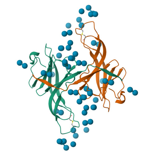
PROTEASE INHIBITOR ECOTIN(1996) Protein Sci 5: 2236-2247
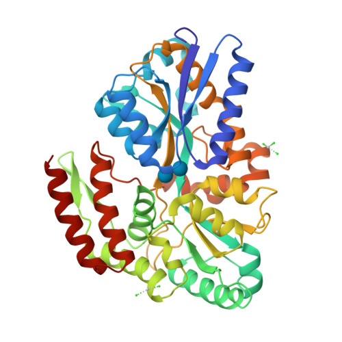
STRUCTURE OF TREHALOSE MALTOSE BINDING PROTEIN FROM THERMOCOCCUS LITORALISDiez, J., Diederichs, K., Greller, G., Horlacher, R., Boos, W., Welte, W. (2001) J Mol Biology 305: 905-915

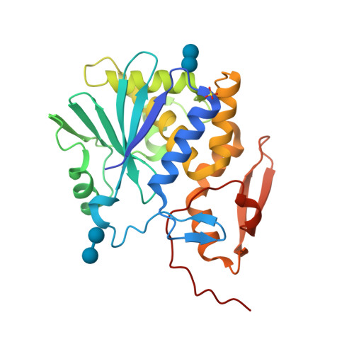
MYCOBACTERIUM TUBERCULOSIS ANTIGEN 85B WITH TREHALOSEAnderson, D.H., Harth, G., Horwitz, M.A., Eisenberg, D., TB Structural Genomics Consortium (TBSGC) (2001) J Mol Biology 307: 671-681
Ricin A-Chain (Recombinant) at 100KTo be published

MALTOPORIN TREHALOSE COMPLEX(1997) J Mol Biology 272: 56-63

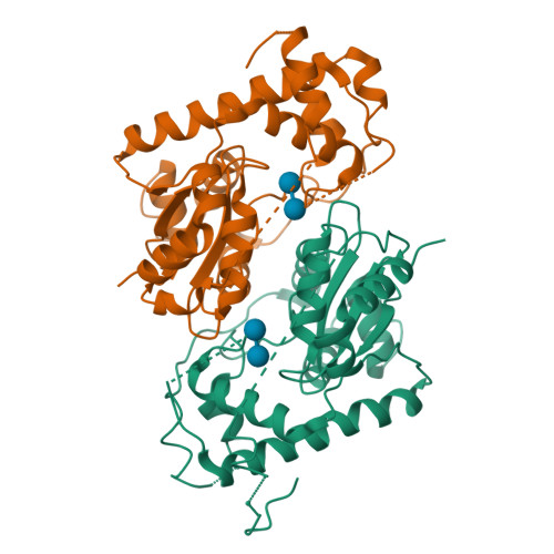
HUMAN GLUCOSAMINE-6-PHOSPHATE DEAMINASE ISOMERASE AT 1.75 A RESOLUTION COMPLEXED WITH N-ACETYL-GLUCOSAMINE-6-PHOSPHATE AND 2-DEOXY-2-AMINO-GLUCITOL-6-PHOSPHATEArreola, R., Valderrama, B., Morante, M.L., Horjales, E. (2003) FEBS Lett 551: 63-70

Comparisions of the Heme-Free and-Bound Crystal Structures of Human Heme Oxygenase-1Lad, L., Schuller, D.J., Friedman, J., Li, H., Shimizu, H., Ortiz de Montellano, P.R., Poulos, T.L. (2003) J Biological Chem 278: 7834-7843
Mycobacterium smegmatis Stf0 Sulfotransferase with TrehaloseMougous, J.D., Petzold, C.J., Senaratne, R.H., Lee, D.H., Akey, D.L., Lin, F.L., Munchel, S.E., Pratt, M.R., Riley, L.W., Leary, J.A., Berger, J.M., Bertozzi, C.R. (2004) Nat Struct Mol Biol 11: 721-729

Crystal Structure of L-lactate dehydrogenase from Cyprinus carpioTo be published
X-ray structure of the sucrose-phosphatase (SPP) from Synechocystis sp.PCC6803 in complex with trehaloseFieulaine, S., Lunn, J.E., Ferrer, J.-L. (2007) Proteins 68: 796-801
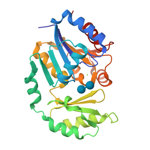
Crystal structure of Deinococcus radiodurans maltooligosyltrehalose trehalohydrolase in complex with trehaloseTimmins, J., Leiros, H.-K.S., Leonard, G., Leiros, I., McSweeney, S. (2005) J Mol Biology 347: 949
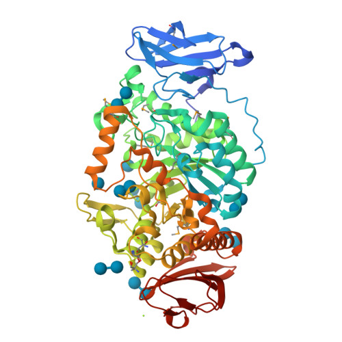
Is radiation damage dependent on the dose-rate used during macromolecular crystallography data collectionLeiros, H.-K.S., Timmins, J., Ravelli, R.B.G., McSweeney, S.M. (2006) Acta Crystallogr D Biol Crystallogr 62: 125

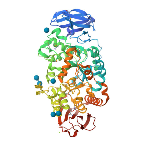
Is radiation damage dependent on the dose-rate used during macromolecular crystallography data collectionLeiros, H.-K.S., Timmins, J., Ravelli, R.B.G., McSweeney, S.M. (2006) Acta Crystallogr D Biol Crystallogr 62: 125

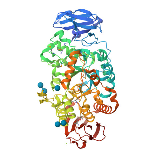
Is radiation damage dependent on the dose-rate used during macromolecular crystallography data collectionLeiros, H.-K.S., Timmins, J., Ravelli, R.B.G., McSweeney, S.M. (2006) Acta Crystallogr D Biol Crystallogr 62: 125
Is radiation damage dependent on the dose-rate used during macromolecular crystallography data collectionLeiros, H.-K.S., Timmins, J., Ravelli, R.B.G., McSweeney, S.M. (2006) Acta Crystallogr D Biol Crystallogr 62: 125
Is radiation damage dependent on the dose-rate used during macromolecular crystallography data collectionLeiros, H.-K.S., Timmins, J., Ravelli, R.B.G., McSweeney, S.M. (2006) Acta Crystallogr D Biol Crystallogr 62: 125
Is radiation damage dependent on the dose-rate used during macromolecular crystallography data collectionLeiros, H.-K.S., Timmins, J., Ravelli, R.B.G., McSweeney, S.M. (2006) Acta Crystallogr D Biol Crystallogr 62: 125
Crystal structure of ConM in complex with trehalose and maltoseCavada, B.S., Azevedo Jr., W.F., Delatorre, P., Rocha, B.A.M., Souza, E.P., Gadelha, C.A.A. (2006) J Struct Biol 154: 280-286
Structure of the complex of C-terminal lobe of bovine lactoferrin with trehalose at 2.0 A resolutionMir, R., Singh, N., Sinha, M., Sharma, S., Bhushan, A., Singh, T.P. To be published

Crystal structure of BphA3 (oxidized form)Senda, M., Kishigami, S., Kimura, S., Ishida, T., Fukuda, M., Senda, T. (2007) J Mol Biology 373: 382-400
Crystal structure of SET/TAF-1beta/INHATMuto, S., Senda, M., Senda, T., Horikoshi, M. (2007) Proc Natl Acad Sci U S A 104: 4285-4290
1 to 25 of 99 Structures Page 1 of 4 Sort by |


