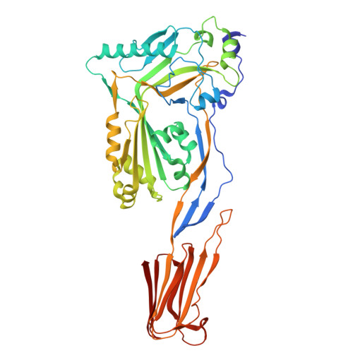
Structure Determination MethodologyScientific Name of Source OrganismMore... Refinement Resolution (Å)Enzyme Classification NameMembrane Protein Annotation | Tsuge, H., Nagahama, M., Nishimura, H., Hisatsune, J., Sakaguchi, Y., Itogawa, Y., Katunuma, N., Sakurai, J. (2003) J Mol Biology 325: 471-483 | Released | 2003-01-14 | | Method | X-RAY DIFFRACTION 1.8 Å | | Organisms | | | Macromolecule | | | Unique Ligands | NAI |
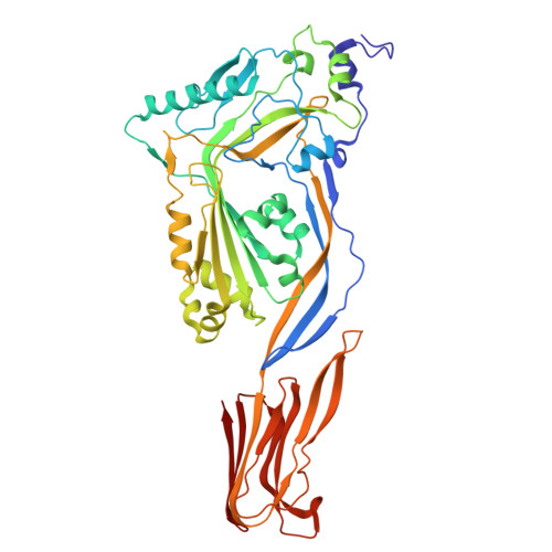
Tsuge, H., Nagahama, M., Nishimura, H., Hisatsune, J., Sakaguchi, Y., Itogawa, Y., Katunuma, N., Sakurai, J. (2003) J Mol Biology 325: 471-483 | Released | 2003-01-14 | | Method | X-RAY DIFFRACTION 2.1 Å | | Organisms | | | Macromolecule | | | Unique Ligands | NDP |
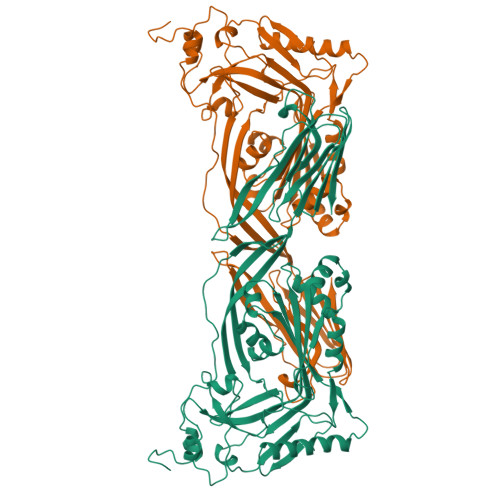
Tempel, W., Liu, Z.-J., Horanyi, P.S., Deng, L., Lee, D., Newton, M.G., Rose, J.P., Ashida, H., Li, S.-C., Li, Y.-T., Wang, B.-C., Southeast Collaboratory for Structural Genomics (SECSG) (2005) Proteins 59: 141-144 | Released | 2004-11-25 | | Method | X-RAY DIFFRACTION 1.82 Å | | Organisms | | | Macromolecule | | | Unique Ligands | CA |
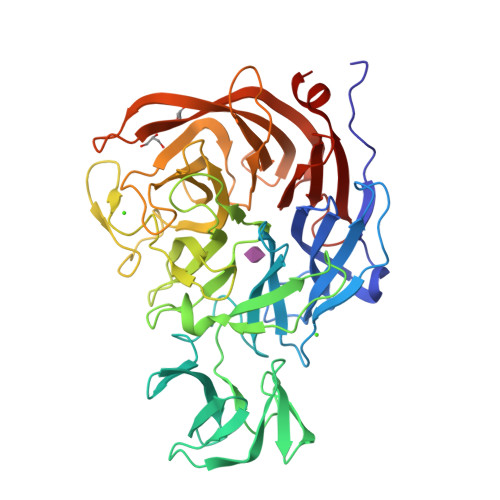
Cole, A.R., Gibert, M., Poppoff, M., Moss, D.S., Titball, R.W., Basak, A.K. (2004) Nat Struct Mol Biol 11: 797 | Released | 2004-08-05 | | Method | X-RAY DIFFRACTION 2.6 Å | | Organisms | | | Macromolecule | | | Unique Ligands | U1 |
Rossocha, M., Schultz-Heienbrok, R., Von Moeller, H., Coleman, J.P., Saenger, W. (2005) Biochemistry 44: 5739 | Released | 2005-03-03 | | Method | X-RAY DIFFRACTION 1.67 Å | | Organisms | | | Macromolecule | | | Unique Ligands | DXC, GOL, TAU |
Rao, F.V., Dorfmueller, H.C., Villa, F., Allwood, M., Eggleston, I.M., van Aalten, D.M.F. (2006) EMBO J 25: 1569-1578 | Released | 2006-02-13 | | Method | X-RAY DIFFRACTION 2.25 Å | | Organisms | | | Macromolecule | | | Unique Ligands | CL, GBL, GOL, SO4, ZN |
Rao, F.V., Dorfmueller, H.C., Villa, F., Allwood, M., Eggleston, I.M., van Aalten, D.M.F. (2006) EMBO J 25: 1569-1578 | Released | 2006-02-13 | | Method | X-RAY DIFFRACTION 2.35 Å | | Organisms | | | Macromolecule | | | Unique Ligands | CL, OAN |
Ficko-Blean, E., Boraston, A.B. (2006) J Biological Chem 281: 37748 | Released | 2006-09-20 | | Method | X-RAY DIFFRACTION 2.4 Å | | Organisms | | | Macromolecule | | | Unique Ligands | CA | | Unique branched monosaccharides | GAL, NDG |
Dorfmueller, H.C., Borodkin, V.S., Schimpl, M., Shepherd, S.M., Shpiro, N.A., van Aalten, D.M.F. (2006) J Am Chem Soc 128: 16484-16485 | Released | 2007-02-13 | | Method | X-RAY DIFFRACTION 2.26 Å | | Organisms | | | Macromolecule | | | Unique Ligands | CL, GSZ |
Ficko-Blean, E., Boraston, A.B. (2006) J Biological Chem 281: 37748 | Released | 2006-10-16 | | Method | X-RAY DIFFRACTION 2.3 Å | | Organisms | | | Macromolecule | | | Unique Ligands | CA | | Unique branched monosaccharides | FUC, GAL, NDG |
Chitayat, S., Gregg, K., Adams, J.J., Ficko-Blean, E., Bayer, E.A., Boraston, A.B., Smith, S.P. (2008) J Mol Biology 375: 20 | Released | 2007-11-06 | | Method | X-RAY DIFFRACTION 2.5 Å | | Organisms | | | Macromolecule | |
|