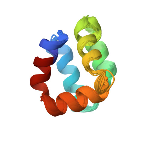
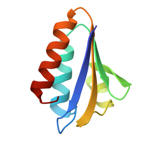
Structure Determination MethodologyScientific Name of Source OrganismMore... Refinement Resolution (Å)Enzyme Classification NameMembrane Protein Annotation | Gonzalez, C., Langdon, G., Bruix, M., Galvez, A., Valdivia, E., Maqueda, M., Rico, M. (2000) Proc Natl Acad Sci U S A 97: 11221 | Released | 2000-10-25 | | Method | SOLUTION NMR | | Organisms | | | Macromolecule | |
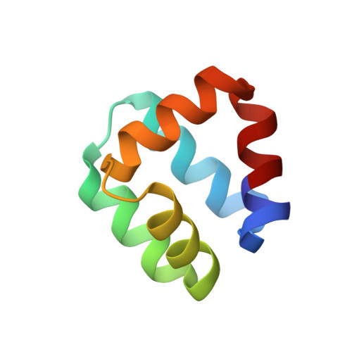
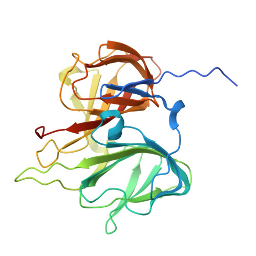
Audette, G.F., Engelmann, R., Hengstenberg, W., Deutscher, J., Hayakawa, K., Quail, J.W., Delbaere, L.T.J. (2000) J Mol Biology 303: 545-553 | Released | 2000-11-22 | | Method | X-RAY DIFFRACTION 1.9 Å | | Organisms | | | Macromolecule | |
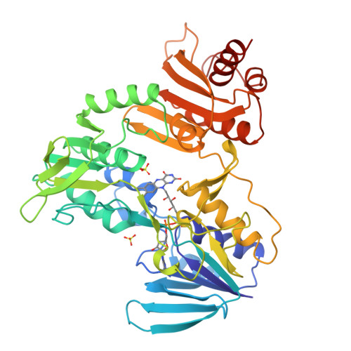
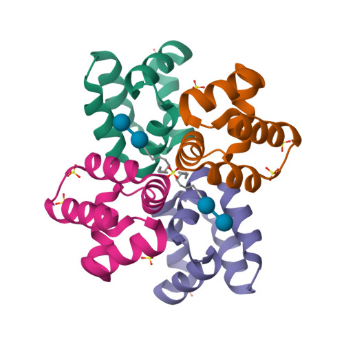
Sanchez-Barrena, M.J., Martinez-Ripoll, M., Galvez, A., Martinez-Bueno, M., Maqueda, M., Cruz, V., Albert, A. (2003) J Mol Biology 334: 541 | Released | 2003-11-20 | | Method | X-RAY DIFFRACTION 1.46 Å | | Organisms | | | Macromolecule | | | Unique Ligands | GOL, SO4 |
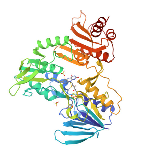
Sanchez-Barrena, M.J., Martinez-Ripoll, M., Galvez, A., Valdivia, E., Maqueda, M., Cruz, V., Albert, A. (2003) J Mol Biology 334: 541 | Released | 2003-11-20 | | Method | X-RAY DIFFRACTION 1.64 Å | | Organisms | | | Macromolecule | | | Unique Ligands | GOL, PO4 |
Sanchez-Barrena, M.J., Martinez-Ripoll, M., Galvez, A., Valdivia, E., Maqueda, M., Cruz, V., Albert, A. (2003) J Mol Biology 334: 541 | Released | 2003-11-20 | | Method | X-RAY DIFFRACTION 2.8 Å | | Organisms | | | Macromolecule | | | Unique Ligands | D10, GOL, SO4 | | Unique branched monosaccharides | GLC |
|