Structure Determination MethodologyScientific Name of Source OrganismRefinement Resolution (Å)Enzyme Classification NameMembrane Protein Annotation | Garcia-Castellanos, R., Mallorqui-Fernandez, G., Marrero, A., Potempa, J., Coll, M., Gomis-Ruth, F.X. (2004) J Biological Chem 279: 17888-17896 | Released | 2004-04-27 | | Method | X-RAY DIFFRACTION 2.8 Å | | Organisms | | | Macromolecule | | | Unique Ligands | K |
Rotella, F.J., Zhang, R.G., Kim, Y., Quartey, P., Collart, F., Joachimiak, A., Midwest Center for Structural Genomics (MCSG) To be published | Released | 2004-11-02 | | Method | X-RAY DIFFRACTION 2.1 Å | | Organisms | | | Macromolecule | |
Kim, Y., Quartey, P., Holzle, D., Collart, F., Joachimiak, A., Midwest Center for Structural Genomics (MCSG) To be published | Released | 2005-09-06 | | Method | X-RAY DIFFRACTION 2.01 Å | | Organisms | | | Macromolecule | | | Unique Ligands | MG |
Marrero, A., Mallorqui-Fernandez, G., Guevara, T., Garcia-Castellanos, R., Gomis-Ruth, F.X. (2006) J Mol Biology 361: 506 | Released | 2006-07-04 | | Method | X-RAY DIFFRACTION 1.8 Å | | Organisms | | | Macromolecule | | | Unique Ligands | GOL, NI |
Patskovsky, Y., Fedorov, E., Toro, R., Sauder, J.M., Smith, D., Freeman, J., Maletic, M., Powell, A., Gheyi, T., Wasserman, S.R., Burley, S.K., Almo, S.C., New York SGX Research Center for Structural Genomics (NYSGXRC) To be published | Released | 2007-02-06 | | Method | X-RAY DIFFRACTION 2.2 Å | | Organisms | | | Macromolecule | | | Unique Ligands | GOL, SO4, ZN |
Fedorov, A.A., Fedorov, E.V., Sauder, J.M., Burley, S.K., Almo, S.C., New York SGX Research Center for Structural Genomics (NYSGXRC) To be published | Released | 2007-04-03 | | Method | X-RAY DIFFRACTION 2.6 Å | | Organisms | | | Macromolecule | | | Unique Ligands | GOL, NA |
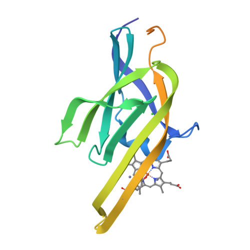
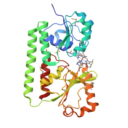
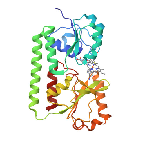
Grigg, J.C., Murphy, M.E. (2007) J Biological Chem 282: 28815-28822 | Released | 2007-07-31 | | Method | X-RAY DIFFRACTION 1.95 Å | | Organisms | | | Macromolecule | | | Unique Ligands | HEM |
Grigg, J.C., Murphy, M.E. (2007) J Biological Chem 282: 28815-28822 | Released | 2007-07-31 | | Method | X-RAY DIFFRACTION 2.15 Å | | Organisms | | | Macromolecule | | | Unique Ligands | HEM |
Bax, B.D., Chan, P.F., Eggleston, D.S., Fosberry, A., Gentry, D.R., Gorrec, F., Giordano, I., Hann, M.M., Hennessy, A., Hibbs, M., Huang, J., Jones, E., Jones, J., Brown, K.K., Lewis, C.J., May, E.W., Singh, O., Spitzfaden, C., Shen, C., Shillings, A., Theobald, A.F., Wohlkonig, A., Pearson, N.D., Gwynn, M.N. (2010) Nature 466: 935 | Released | 2010-08-04 | | Method | X-RAY DIFFRACTION 3.1 Å | | Organisms | | | Macromolecule | | | Unique Ligands | CA |
Bax, B.D., Chan, P.F., Eggleston, D.S., Fosberry, A., Gentry, D.R., Gorrec, F., Giordano, I., Hann, M.M., Hennessy, A., Hibbs, M., Huang, J., Jones, E., Jones, J., Brown, K.K., Lewis, C.J., May, E.W., Singh, O., Spitzfaden, C., Shen, C., Shillings, A., Theobald, A.F., Wohlkonig, A., Pearson, N.D., Gwynn, M.N. (2010) Nature 466: 935 | Released | 2010-08-04 | | Method | X-RAY DIFFRACTION 2.98 Å | | Organisms | | | Macromolecule | |
Bax, B.D., Chan, P.F., Eggleston, D.S., Fosberry, A., Gentry, D.R., Gorrec, F., Giordano, I., Hann, M.M., Hennessy, A., Hibbs, M., Huang, J., Jones, E., Jones, J., Brown, K.K., Lewis, C.J., May, E.W., Singh, O., Spitzfaden, C., Shen, C., Shillings, A., Theobald, A.F., Wohlkonig, A., Pearson, N.D., Gwynn, M.N. (2010) Nature 466: 935 | Released | 2010-08-04 | | Method | X-RAY DIFFRACTION 2.1 Å | | Organisms | | | Macromolecule | | | Unique Ligands | MN, RXV |
Bax, B.D., Chan, P., Eggleston, D.S., Fosberry, A., Gentry, D.R., Gorrec, F., Giordano, I., Hann, M.M., Hennessy, A., Hibbs, M., Huang, J., Jones, E., Jones, J., Brown, K.K., Lewis, C.J., May, E., Singh, O., Spitzfaden, C., Shen, C., Shillings, A., Theobald, A., Wohlkonig, A., Pearson, N.D., Gwynn, M.N. (2010) Nature 466: 935 | Released | 2010-08-25 | | Method | X-RAY DIFFRACTION 3.35 Å | | Organisms | | | Macromolecule | | | Unique Ligands | CPF, MN |
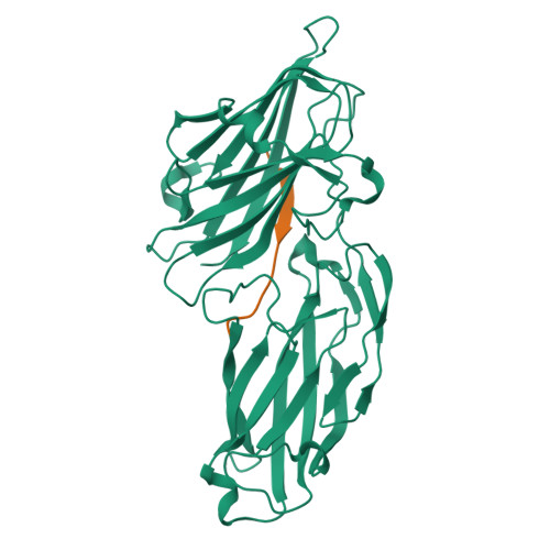
Ganesh, V.K. To be published | Released | 2011-05-04 | | Method | X-RAY DIFFRACTION 2.6 Å | | Organisms | | | Macromolecule | |
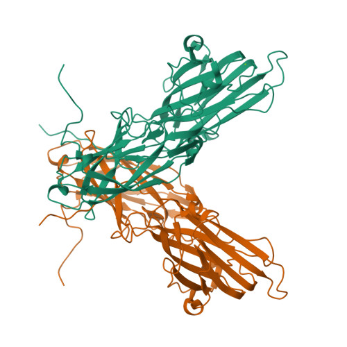
Ganesh, V.K. To be published | Released | 2011-05-04 | | Method | X-RAY DIFFRACTION 2.5 Å | | Organisms | | | Macromolecule | |
Ganesh, V.K., Barbu, E.M., Deivanayagam, C.C.S., Le, B., Anderson, A.S., Matsuka, Y., Lin, S.L., Foster, T.F., Narayana, S.V.L., Hook, M. To be published | Released | 2011-05-04 | | Method | X-RAY DIFFRACTION 2.45 Å | | Organisms | | | Macromolecule | | | Unique Ligands | MG |
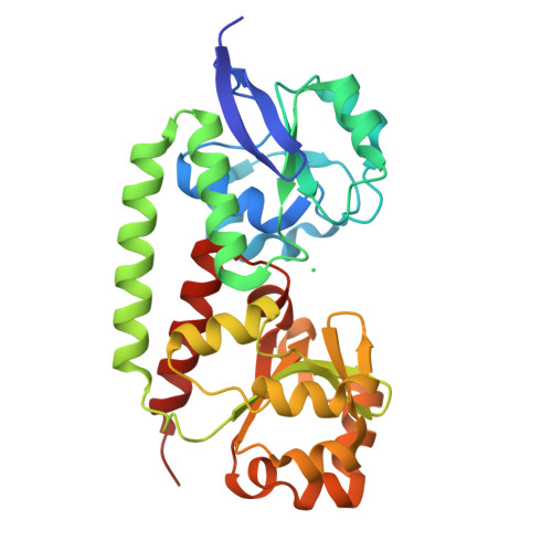
Grigg, J.C., Murphy, M.E.P. (2009) Mol Microbiol 72: 947-963 | Released | 2009-05-26 | | Method | X-RAY DIFFRACTION 1.6 Å | | Organisms | | | Macromolecule | | | Unique Ligands | CL |
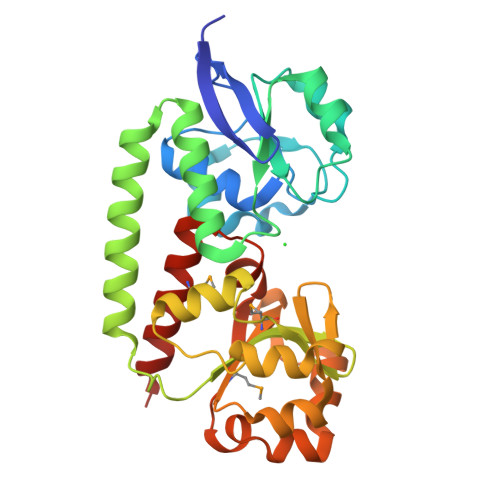
Grigg, J.C., Murphy, M.E.P. (2009) Mol Microbiol 72: 947-963 | Released | 2009-05-26 | | Method | X-RAY DIFFRACTION 1.35 Å | | Organisms | | | Macromolecule | | | Unique Ligands | CL |
Patskovsky, Y., Toro, R., Dickey, M., Chang, S., Sauder, J.M., Burley, S.K., Almo, S.C., New York SGX Research Center for Structural Genomics (NYSGXRC) To be published | Released | 2009-08-25 | | Method | X-RAY DIFFRACTION 2.3 Å | | Organisms | | | Macromolecule | |
Fan, Y., Xu, X., Cui, H., Ng, J., Savchenko, A., Joachimiak, A., Edwards, A., Midwest Center for Structural Genomics (MCSG) To be published | Released | 2009-09-15 | | Method | X-RAY DIFFRACTION 1.5 Å | | Organisms | | | Macromolecule | |
|