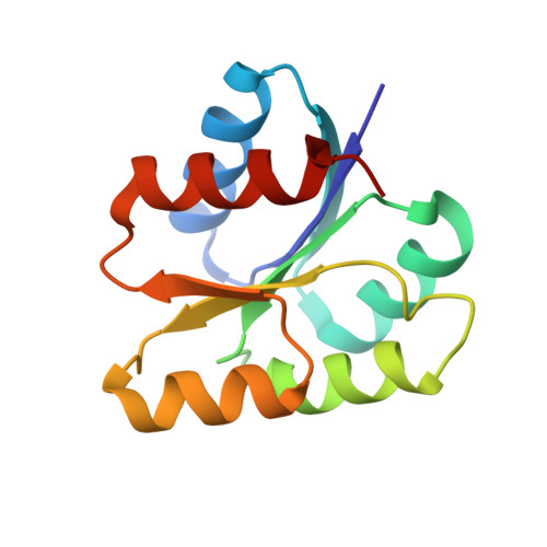
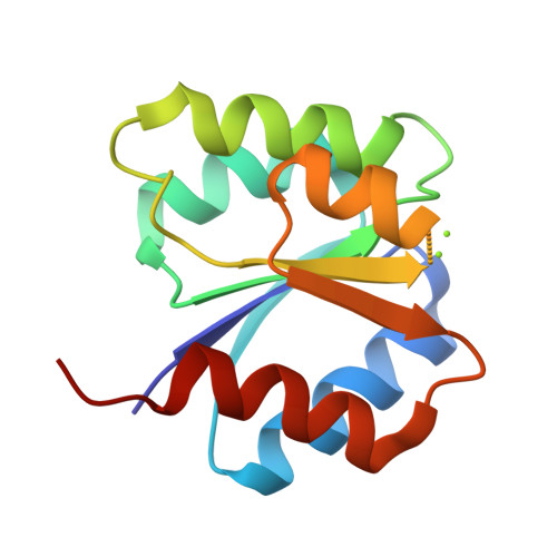
Structure Determination MethodologyScientific Name of Source OrganismRefinement Resolution (Å)Enzyme Classification Name | Guillet, V., Ohta, N., Cabantous, S., Newton, A., Samama, J.-P., Structural Proteomics in Europe (SPINE) (2002) J Biological Chem 277: 42003-42010 | Released | 2002-11-15 | | Method | X-RAY DIFFRACTION 1.6 Å | | Organisms | | | Macromolecule | |
Guillet, V., Ohta, N., Cabantous, S., Newton, A., Samama, J.-P., Structural Proteomics in Europe (SPINE) (2002) J Biological Chem 277: 42003-42010 | Released | 2002-11-15 | | Method | X-RAY DIFFRACTION 1.87 Å | | Organisms | | | Macromolecule | |
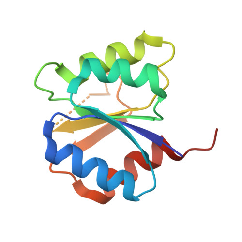
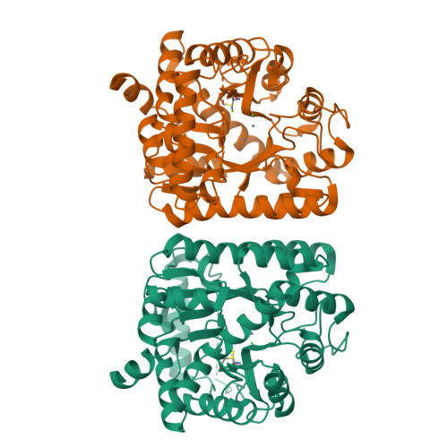
Guillet, V., Ohta, N., Cabantous, S., Newton, A., Samama, J.-P., Structural Proteomics in Europe (SPINE) (2002) J Biological Chem 277: 42003-42010 | Released | 2002-12-04 | | Method | X-RAY DIFFRACTION 1.6 Å | | Organisms | | | Macromolecule | | | Unique Ligands | MN |
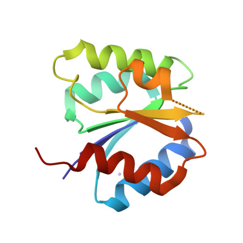
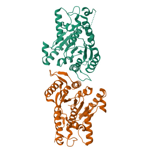
Guillet, V., Ohta, N., Cabantous, S., Newton, A., Samama, J.-P., Structural Proteomics in Europe (SPINE) (2002) J Biological Chem 277: 42003-42010 | Released | 2002-12-04 | | Method | X-RAY DIFFRACTION 2 Å | | Organisms | | | Macromolecule | | | Unique Ligands | MN |
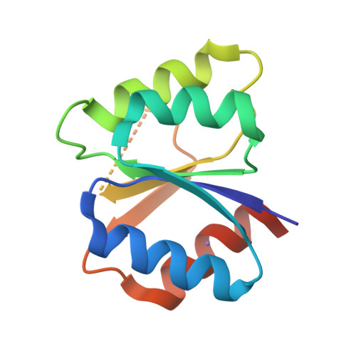
Guillet, V., Ohta, N., Cabantous, S., Newton, A., Samama, J.-P., Structural Proteomics in Europe (SPINE) (2002) J Biological Chem 277: 42003-42010 | Released | 2002-12-04 | | Method | X-RAY DIFFRACTION 1.41 Å | | Organisms | | | Macromolecule | | | Unique Ligands | MG |
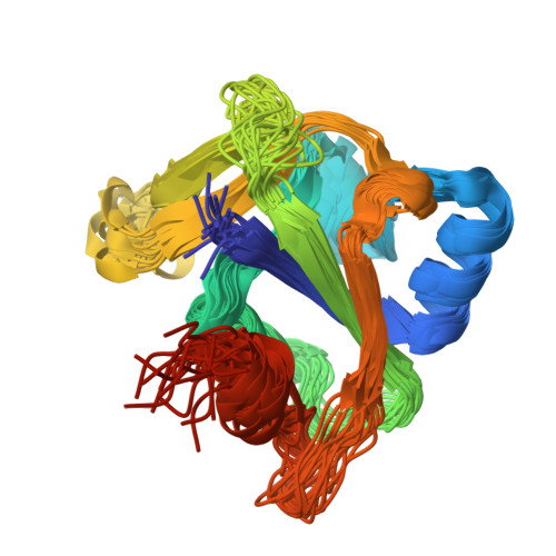
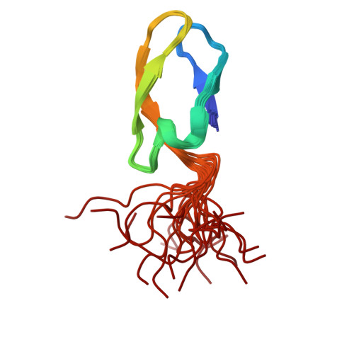
Shen, Y., Atreya, H.S., Acton, T., Xiao, R., Montelione, G.T., Szyperski, T., Northeast Structural Genomics Consortium (NESG) (2005) Proteins 58: 747-750 | Released | 2005-01-04 | | Method | SOLUTION NMR | | Organisms | | | Macromolecule | |
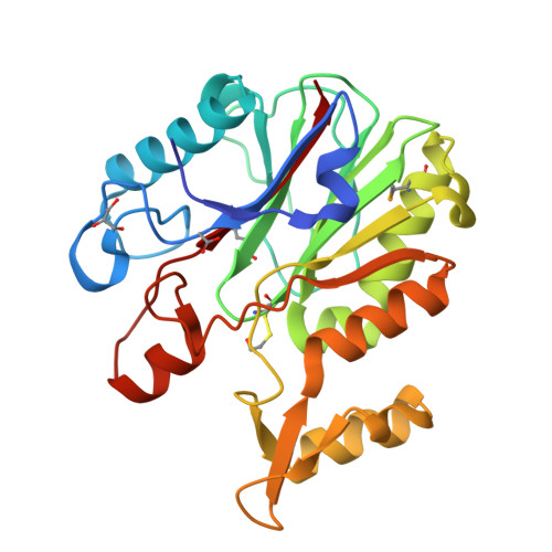
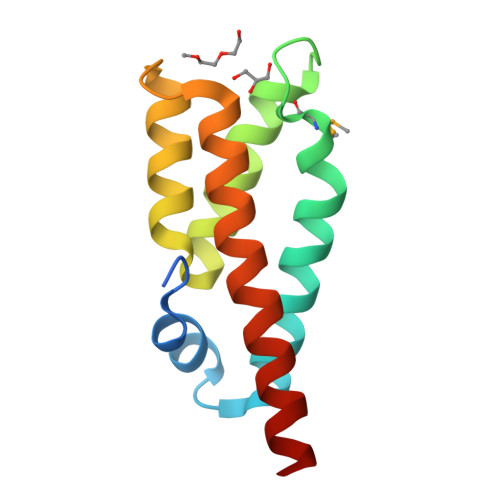
Krishnamurthy, N.R., Kumaran, D., Swaminathan, S., Burley, S.K., New York SGX Research Center for Structural Genomics (NYSGXRC) To be published | Released | 2004-09-21 | | Method | X-RAY DIFFRACTION 1.75 Å | | Organisms | | | Macromolecule | | | Unique Ligands | ACT, BME, GOL |
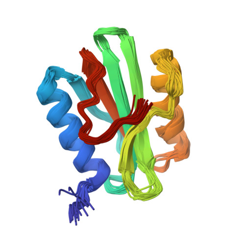
Aramini, J.M., Rossi, P., Moseley, H.N.B., Wang, D., Nwosu, C., Cunningham, K., Ma, L., Xiao, R., Liu, J., Baran, M.C., Swapna, G.V.T., Acton, T.B., Rost, B., Montelione, G.T., Northeast Structural Genomics Consortium (NESG) To be published | Released | 2007-07-03 | | Method | SOLUTION NMR | | Organisms | | | Macromolecule | |
Shi, C., Fricke, P., Lin, L., Chevelkov, V., Wegstroth, M., Giller, K., Becker, S., Thanbichler, M., Lange, A. (2015) Sci Adv 1: e1501087-e1501087 | Released | 2015-12-16 | | Method | SOLID-STATE NMR | | Organisms | | | Macromolecule | |
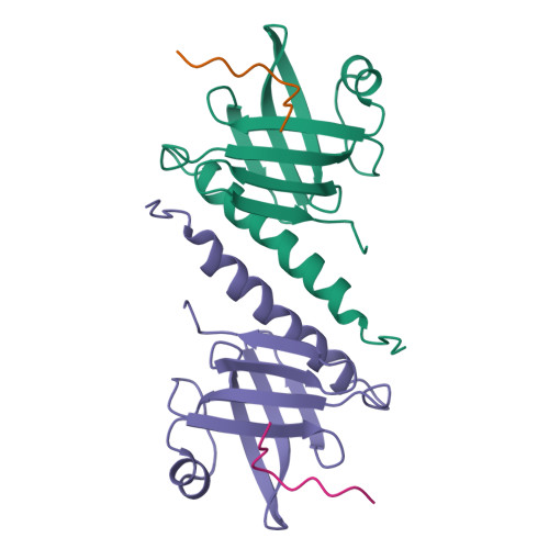
Seetharaman, J., Su, M., Wang, D., Fang, Y., Cunningham, K., Ma, L., Xiao, R., Liu, J., Baran, M.C., Acton, T.B., Rost, B., Montelione, G.T., Hunt, J.F., Tong, L., Northeast Structural Genomics Consortium (NESG) To be published | Released | 2006-12-12 | | Method | X-RAY DIFFRACTION 1.9 Å | | Organisms | | | Macromolecule | |
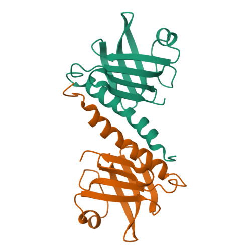
Seetharaman, J., Su, M., Wang, D., Fang, Y., Cunningham, K., Ma, L., Xiao, R., Liu, J., Baran, M.C., Acton, T.B., Rost, B., Montelione, G.T., Hunt, J.F., Tong, L., Northeast Structural Genomics Consortium (NESG) To be published | Released | 2006-12-12 | | Method | X-RAY DIFFRACTION 1.8 Å | | Organisms | | | Macromolecule | |
Patskovsky, Y., Bonanno, J., Sridhar, V., Sauder, J.M., Freeman, J., Powell, A., Koss, J., Groshong, C., Gheyi, T., Wasserman, S.R., Raushel, F., Burley, S.K., Almo, S.C., New York SGX Research Center for Structural Genomics (NYSGXRC) To be published | Released | 2007-05-29 | | Method | X-RAY DIFFRACTION 2.34 Å | | Organisms | | | Macromolecule | | | Unique Ligands | K |
Fedorov, A.A., Fedorov, E.V., Toro, R., Sauder, J.M., Burley, S.K., Almo, S.C., New York SGX Research Center for Structural Genomics (NYSGXRC) To be published | Released | 2007-10-02 | | Method | X-RAY DIFFRACTION 2 Å | | Organisms | | | Macromolecule | | | Unique Ligands | CL, ZN |
|