Structure Determination MethodologyScientific Name of Source OrganismRefinement Resolution (Å)Enzyme Classification Name | Ahn, S., Milner, A.J., Futterer, K., Konopka, M., Ilias, M., Young, T.W., White, S.A. (2001) J Mol Biology 313: 797-811 | Released | 2001-10-31 | | Method | X-RAY DIFFRACTION 1.5 Å | | Organisms | | | Macromolecule | | | Unique Ligands | GOL, MN, SO4 |
Fabrichniy, I.P., Lehtio, L., Salminen, A., Baykov, A.A., Lahti, R., Goldman, A. (2004) Biochemistry 43: 14403-14411 | Released | 2004-11-23 | | Method | X-RAY DIFFRACTION 2.05 Å | | Organisms | | | Macromolecule | | | Unique Ligands | CL, SO4, ZN |
Langley, D.B. (2008) J Mol Biology 377: 104-116 | Released | 2008-03-11 | | Method | X-RAY DIFFRACTION 1.85 Å | | Organisms | | | Macromolecule | | | Unique Ligands | SO4 |
Langley, D.B. (2008) J Mol Biology 377: 104-116 | Released | 2008-03-11 | | Method | X-RAY DIFFRACTION 1.61 Å | | Organisms | | | Macromolecule | | | Unique Ligands | NGT |
Langley, D.B. (2008) J Mol Biology 377: 104-116 | Released | 2008-03-11 | | Method | X-RAY DIFFRACTION 1.56 Å | | Organisms | | | Macromolecule | | | Unique Ligands | ACY |
Forsgren, N., Persson, K. (2009) Protein Sci 18: 1896 | Released | 2009-07-28 | | Method | X-RAY DIFFRACTION 2.3 Å | | Organisms | | | Macromolecule | | | Unique Ligands | CA, GOL, K |
Forsgren, N., Persson, K. (2010) J Mol Biology 397: 740 | Released | 2010-02-16 | | Method | X-RAY DIFFRACTION 1.5 Å | | Organisms | | | Macromolecule | | | Unique Ligands | CA |
Forsgren, N., Persson, K. (2010) J Mol Biology 397: 740 | Released | 2010-02-16 | | Method | X-RAY DIFFRACTION 1.7 Å | | Organisms | | | Macromolecule | | | Unique Ligands | CA, MG |
Stoll, K.E., Draper, W.E., Kliegman, J.I., Golynskiy, M.V., Brew-Appiah, R.A.T., Brown, H.K., Breyer, W.A., Jakubovics, N.S., Jenkinson, H.F., Brennan, R.B., Cohen, S.M., Glasfeld, A. (2009) Biochemistry 48: 10308-10320 | Released | 2009-06-23 | | Method | X-RAY DIFFRACTION 2.7 Å | | Organisms | | | Macromolecule | | | Unique Ligands | SO4 |
Stoll, K.E., Draper, W.E., Kliegman, J.I., Golynskiy, M.V., Brew-Appiah, R.A.T., Brown, H.K., Breyer, W.A., Jakubovics, N.S., Jenkinson, H.F., Brennan, R.B., Cohen, S.M., Glasfeld, A. (2009) Biochemistry 48: 10308-10320 | Released | 2009-06-23 | | Method | X-RAY DIFFRACTION 2.8 Å | | Organisms | | | Macromolecule | | | Unique Ligands | CD, SO4 |
Stoll, K.E., Draper, W.E., Kliegman, J.I., Golynskiy, M.V., Brew-Appiah, R.A.T., Brown, H.K., Breyer, W.A., Jakubovics, N.S., Jenkinson, H.F., Brennan, R.B., Cohen, S.M., Glasfeld, A. (2009) Biochemistry 48: 10308-10320 | Released | 2009-06-23 | | Method | X-RAY DIFFRACTION 2.9 Å | | Organisms | | | Macromolecule | | | Unique Ligands | SO4, ZN |
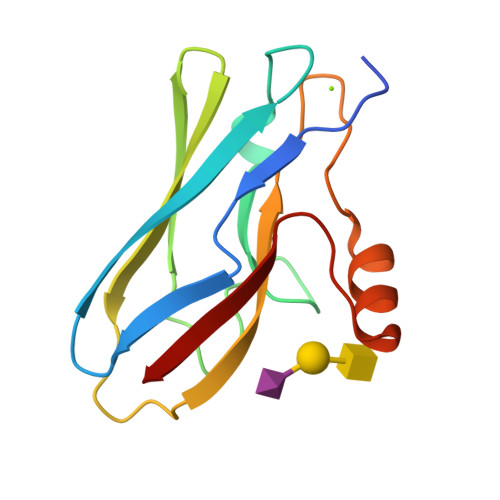
Pyburn, T.M. (2011) PLoS Pathog 7: e1002112-e1002112 | Released | 2011-08-10 | | Method | X-RAY DIFFRACTION 1.9 Å | | Organisms | | | Macromolecule | | | Unique Ligands | CA, GOL, HDZ |
Nylander, A., Svensater, G., Senadheera, D.B., Cvitkovitch, D.G., Davies, J.R., Persson, K. (2013) PLoS One 8: e63768-e63768 | Released | 2013-06-05 | | Method | X-RAY DIFFRACTION 2.09 Å | | Organisms | | | Macromolecule | | | Unique Ligands | ACT, GOL, NA, SO4 |
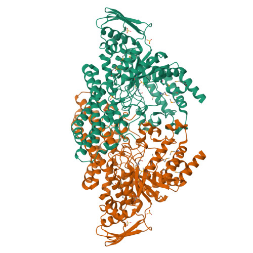
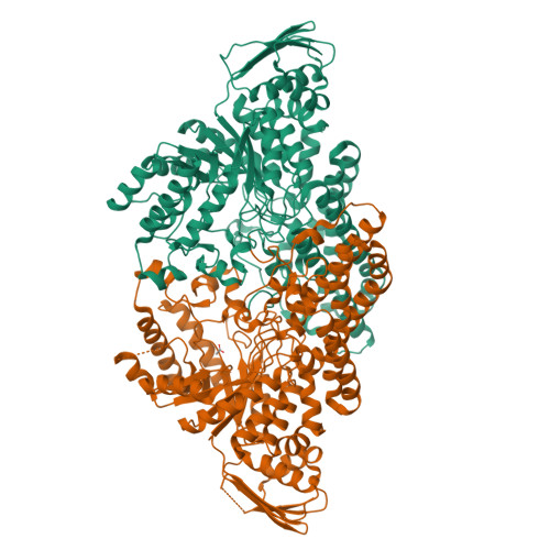
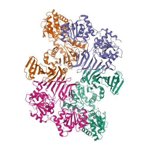
Chen, Y., Rapoport, T.A. (2016) Proc Natl Acad Sci U S A 113: E1190-E1199 | Released | 2016-03-02 | | Method | X-RAY DIFFRACTION 2.92 Å | | Organisms | | | Macromolecule | | | Unique Ligands | MG |
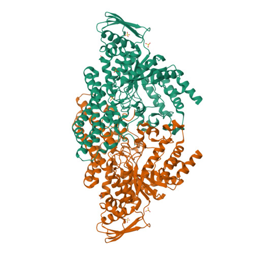
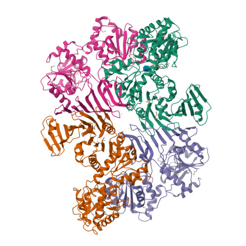
Chen, Y., Rapoport, T.A. (2016) Proc Natl Acad Sci U S A 113: E1190-E1199 | Released | 2016-03-02 | | Method | X-RAY DIFFRACTION 3.84 Å | | Organisms | | | Macromolecule | | | Unique Ligands | MG, NAG, UDP |
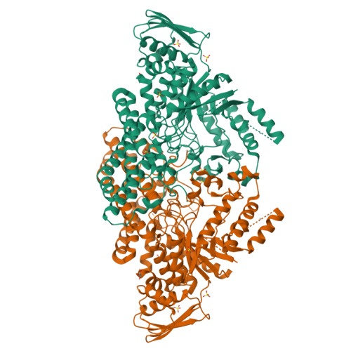
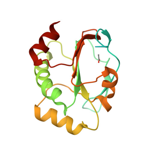
Stogios, P.J., Evdokimova, E., Wawrzak, Z., Yim, V., Savchenko, A., Anderson, W.F., Center for Structural Genomics of Infectious Diseases (CSGID) To be published | Released | 2017-02-15 | | Method | X-RAY DIFFRACTION 1.62 Å | | Organisms | | | Macromolecule | | | Unique Ligands | ACT |
|