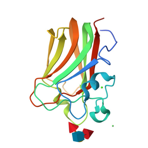
Structure Determination MethodologyScientific Name of Source OrganismRefinement Resolution (Å) | Feil, S.C. (2012) Structure 20: 248-258 | Released | 2010-12-29 | | Method | X-RAY DIFFRACTION 1.91 Å | | Organisms | | | Macromolecule | | | Unique Ligands | CA, GOL, NI |
Feil, S.C. (2012) Structure 20: 248-258 | Released | 2010-12-29 | | Method | X-RAY DIFFRACTION 2.01 Å | | Organisms | | | Macromolecule | | | Unique Ligands | CA, NI | | Unique branched monosaccharides | FUC, GAL, NAG |
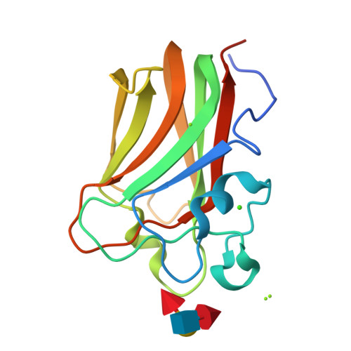
Feil, S.C. (2012) Structure 20: 248-258 | Released | 2010-12-29 | | Method | X-RAY DIFFRACTION 1.9 Å | | Organisms | | | Macromolecule | | | Unique Ligands | CA, FUC, NI |
Feil, S.C. (2012) Structure 20: 248-258 | Released | 2010-12-29 | | Method | X-RAY DIFFRACTION 2 Å | | Organisms | | | Macromolecule | | | Unique Ligands | CA, NI | | Unique branched monosaccharides | FUC, GAL, NDG |
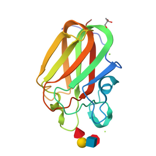
Feil, S.C. (2012) Front Immunol 3: 330-330 | Released | 2012-11-21 | | Method | X-RAY DIFFRACTION 1.6 Å | | Organisms | | | Macromolecule | | | Unique Ligands | CA, GOL, MG | | Unique branched monosaccharides | FUC, GAL, NAG |
Feil, S.C. (2012) Front Immunol 3: 330-330 | Released | 2012-11-21 | | Method | X-RAY DIFFRACTION 1.6 Å | | Organisms | | | Macromolecule | | | Unique Ligands | CA, MG | | Unique branched monosaccharides | FUC, GAL, NAG |
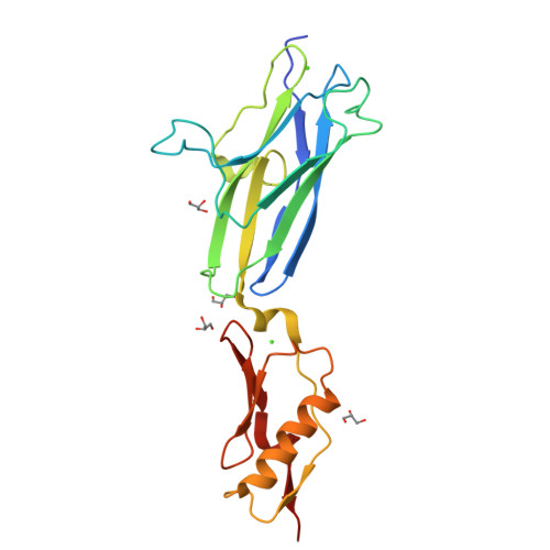
Iverson, T.M. (2022) Nat Commun 13: 2753-2753 | Released | 2020-02-19 | | Method | X-RAY DIFFRACTION 1.6 Å | | Organisms | | | Macromolecule | | | Unique Ligands | ACT, CA, GOL |
|