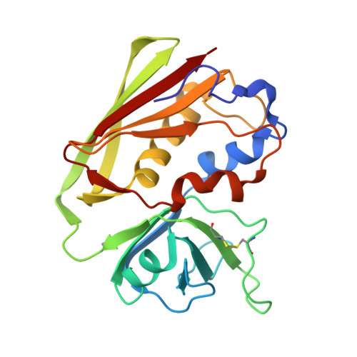
Structure Determination MethodologyScientific Name of Source OrganismMore... Refinement Resolution (Å)Enzyme Classification NameMembrane Protein Annotation | Roussel, A., Baker, E.N. (1997) Nat Struct Biol 4: 635-643 | Released | 1998-04-29 | | Method | X-RAY DIFFRACTION 2.4 Å | | Organisms | | | Macromolecule | |
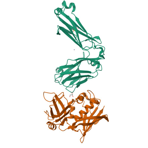
Sundberg, E., Jardetzky, T. (1999) Nat Struct Biol 6: 123-129 | Released | 1998-10-14 | | Method | X-RAY DIFFRACTION 1.85 Å | | Organisms | | | Macromolecule | |
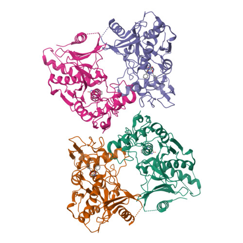
Kagawa, T.F., Cooney, J.C., Baker, H.M., McSweeney, S., Liu, M., Gubba, S., Musser, J.M., Baker, E.N. (2000) Proc Natl Acad Sci U S A 97: 2235-2240 | Released | 2000-03-01 | | Method | X-RAY DIFFRACTION 1.6 Å | | Organisms | | | Macromolecule | | | Unique Ligands | SO4 |
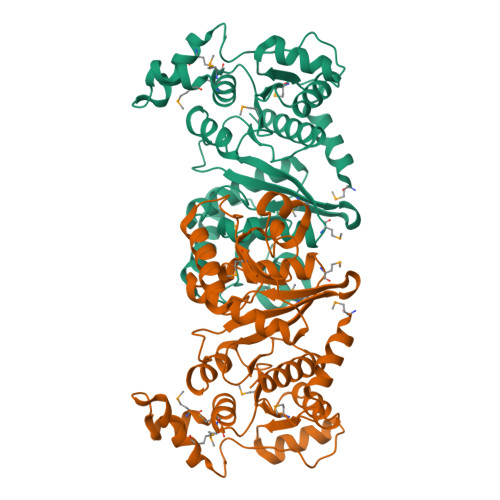
Campbell, R.E., Mosimann, S.C., van de Rijn, I., Tanner, M.E., Strynadka, N.C.J. (2000) Biochemistry 39: 7012-7023 | Released | 2000-05-31 | | Method | X-RAY DIFFRACTION 2.31 Å | | Organisms | | | Macromolecule | | | Unique Ligands | GOL, NAD, SO4, UDX |
Campbell, R.E., Mosimann, S.C., van de Rijn, I., Tanner, M.E., Strynadka, N.C.J. (2000) Biochemistry 39: 7012-7023 | Released | 2000-05-31 | | Method | X-RAY DIFFRACTION 1.8 Å | | Organisms | | | Macromolecule | | | Unique Ligands | GOL, NAI, SO4, UGA |
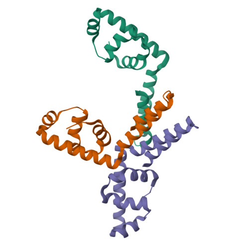
Arcus, V.L., Proft, T., Sigrell, J.A., Baker, H.M., Fraser, J.D., Baker, E.N. (2000) J Mol Biology 299: 157-168 | Released | 2000-05-10 | | Method | X-RAY DIFFRACTION 1.9 Å | | Organisms | | | Macromolecule | |
Arcus, V.L., Proft, T., Sigrell, J.A., Baker, H.M., Fraser, J.D., Baker, E.N. (2000) J Mol Biology 299: 157-168 | Released | 2000-04-26 | | Method | X-RAY DIFFRACTION 1.9 Å | | Organisms | | | Macromolecule | |
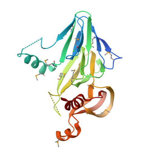
Arcus, V.L., Proft, T., Sigrell, J.A., Baker, H.M., Fraser, J.D., Baker, E.N. (2000) J Mol Biology 299: 157-168 | Released | 2000-04-26 | | Method | X-RAY DIFFRACTION 1.68 Å | | Organisms | | | Macromolecule | | | Unique Ligands | K, PO4, ZN |
Arcus, V.L., Proft, T., Sigrell, J.A., Baker, H.M., Fraser, J.D., Baker, E.N. (2000) J Mol Biology 299: 157-168 | Released | 2000-04-26 | | Method | X-RAY DIFFRACTION 2.5 Å | | Organisms | | | Macromolecule | | | Unique Ligands | ZN |
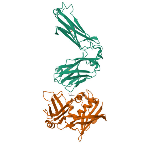
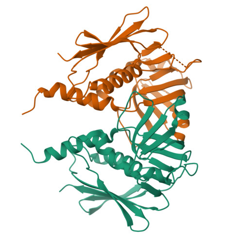
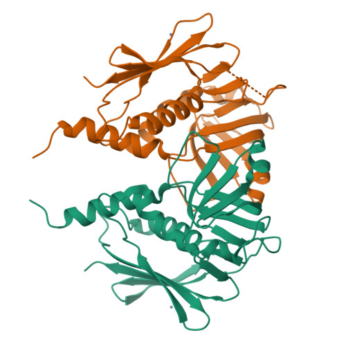
Sundberg, E.J., Li, H., Llera, A.S., McCormick, J.K., Tormo, J., Karjalainen, K., Schlievert, P.M., Mariuzza, R.A. (2002) Structure 10: 687-699 | Released | 2002-06-07 | | Method | X-RAY DIFFRACTION 3 Å | | Organisms | | | Macromolecule | |
Kuzin, A., Lee, I., Edstrom, W., Xiao, R., Acton, T., Rost, B., Montelione, G., Hunt, J., Tong, L., Northeast Structural Genomics Consortium (NESG) (2003) To be published | Released | 2003-12-02 | | Method | X-RAY DIFFRACTION 1.7 Å | | Organisms | | | Macromolecule | | | Unique Ligands | CA |
Xu, Q.S., Shin, D.H., Pufan, R., Yokota, H., Kim, R., Kim, S.H., Berkeley Structural Genomics Center (BSGC) (2004) Proteins 55: 479-481 | Released | 2004-04-13 | | Method | X-RAY DIFFRACTION 2.7 Å | | Organisms | | | Macromolecule | |
Oganesyan, V., Pufan, R., DeGiovanni, A., Yokota, H., Kim, R., Kim, S.-H., Berkeley Structural Genomics Center (BSGC) (2004) Acta Crystallogr D Biol Crystallogr 60: 1266-1271 | Released | 2004-06-29 | | Method | X-RAY DIFFRACTION 2.31 Å | | Organisms | | | Macromolecule | |
Liu, J., Oganesyan, N., Shin, D.-H., Jancarik, J., Pufan, R., Yokota, H., Kim, R., Kim, S.-H., Berkeley Structural Genomics Center (BSGC) (2005) Proteins 59: 875-881 | Released | 2004-08-24 | | Method | X-RAY DIFFRACTION 2.3 Å | | Organisms | | | Macromolecule | | | Unique Ligands | ZN |
Baker, H.M., Proft, T., Webb, P.D., Arcus, V.L., Fraser, J.D., Baker, E.N. (2004) J Biological Chem 279: 38571-38576 | Released | 2004-08-03 | | Method | X-RAY DIFFRACTION 1.75 Å | | Organisms | | | Macromolecule | |
Baker, H.M., Proft, T., Webb, P.D., Arcus, V.L., Fraser, J.D., Baker, E.N. (2004) J Biological Chem 279: 38571-38576 | Released | 2004-08-17 | | Method | X-RAY DIFFRACTION 2 Å | | Organisms | | | Macromolecule | | | Unique Ligands | ZN |
|