1EO2

CRYSTAL STRUCTURE OF ACINETOBACTER SP. ADP1 PROTOCATECHUATE 3,4-DIOXYGENASE


Experimental Data Snapshot

  • Method: X-RAY DIFFRACTION
  • Resolution: 2.25 Å
  • R-Value Free: 0.218 
  • R-Value Work: 0.176 
  • R-Value Observed: 0.176 

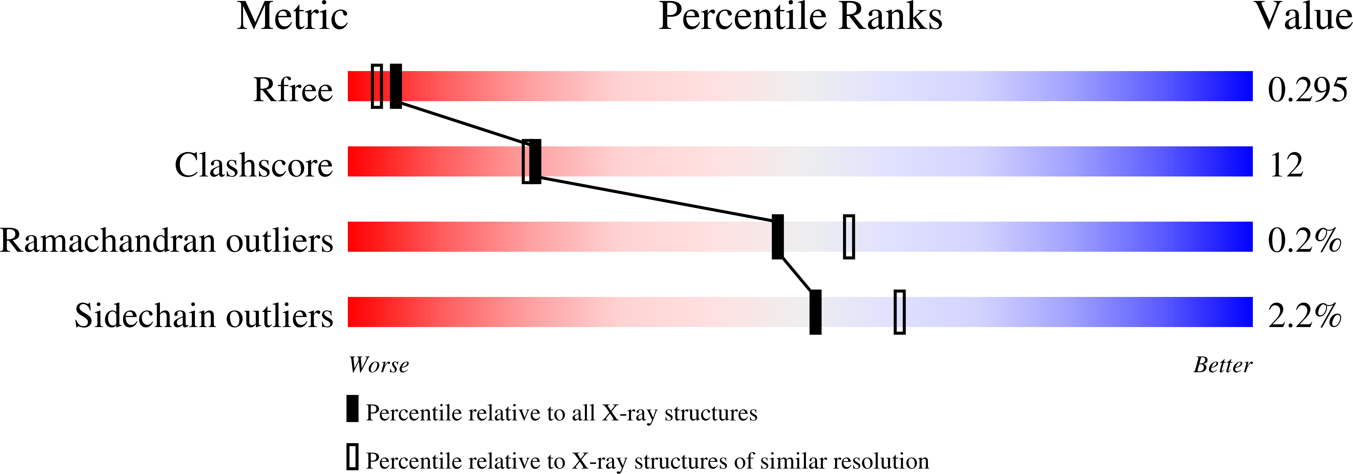
wwPDB Validation   3D Report Full Report


This is version 1.5 of the entry. See complete history


Literature

Structure of Acinetobacter strain ADP1 protocatechuate 3, 4-dioxygenase at 2.2 A resolution: implications for the mechanism of an intradiol dioxygenase.

Vetting, M.W.D'Argenio, D.A.Ornston, L.N.Ohlendorf, D.H.

(2000) Biochemistry 39: 7943-7955

  • DOI: https://doi.org/10.1021/bi000151e
  • Primary Citation of Related Structures:  
    1EO2, 1EO9, 1EOA, 1EOB, 1EOC

  • PubMed Abstract: 

    The crystal structures of protocatechuate 3,4-dioxygenase from the soil bacteria Acinetobacterstrain ADP1 (Ac 3,4-PCD) have been determined in space group I23 at pH 8.5 and 5.75. In addition, the structures of Ac 3,4-PCD complexed with its substrate 3, 4-dihydroxybenzoic acid (PCA), the inhibitor 4-nitrocatechol (4-NC), or cyanide (CN(-)) have been solved using native phases. The overall tertiary and quaternary structures of Ac 3,4-PCD are similar to those of the same enzyme from Pseudomonas putida[Ohlendorf et al. (1994) J. Mol. Biol. 244, 586-608]. At pH 8.5, the catalytic non-heme Fe(3+) is coordinated by two axial ligands, Tyr447(OH) (147beta) and His460(N)(epsilon)(2) (160beta), and three equatorial ligands, Tyr408(OH) (108beta), His462(N)(epsilon)(2) (162beta), and a hydroxide ion (d(Fe-OH) = 1.91 A) in a distorted bipyramidal geometry. At pH 5.75, difference maps suggest a sulfate binds to the Fe(3+) in an equatorial position and the hydroxide is shifted [d(Fe-OH) = 2.3 A] yielding octahedral geometry for the active site Fe(3+). This change in ligation geometry is concomitant with a shift in the optical absorbance spectrum of the enzyme from lambda(max) = 450 nm to lambda(max) = 520 nm. Binding of substrate or 4-NC to the Fe(3+) is bidentate with the axial ligand Tyr447(OH) (147beta) dissociating. The structure of the 4-NC complex supports the view that resonance delocalization of the positive character of the nitrogen prevents substrate activation. The cyanide complex confirms previous work that protocatechuate 3,4-dioxygenases have three coordination sites available for binding by exogenous substrates. A significant conformational change extending away from the active site is seen in all structures when compared to the native enzyme at pH 8.5. This conformational change is discussed in its relevance to enhancing catalysis in protocatechuate 3,4-dioxygenases.


  • Organizational Affiliation

    Center of Metals in Biocatalysis and Department of Biochemistry, Molecular Biology and Biophysics, 6-155 Jackson Hall, 321 Church St. S.E., University of Minnesota Medical School, Minneapolis, Minnesota 55455-0347, USA.


Macromolecules
Find similar proteins by:  (by identity cutoff)  |  3D Structure
Entity ID: 1
MoleculeChains Sequence LengthOrganismDetailsImage
PROTOCATECHUATE 3,4-DIOXYGENASE ALPHA CHAIN209Acinetobacter baylyi ADP1Mutation(s): 0 
EC: 1.13.11.3
UniProt
Find proteins for P20371 (Acinetobacter baylyi (strain ATCC 33305 / BD413 / ADP1))
Explore P20371 
Go to UniProtKB:  P20371
Entity Groups  
Sequence Clusters30% Identity50% Identity70% Identity90% Identity95% Identity100% Identity
UniProt GroupP20371
Sequence Annotations
Expand
  • Reference Sequence
Find similar proteins by:  (by identity cutoff)  |  3D Structure
Entity ID: 2
MoleculeChains Sequence LengthOrganismDetailsImage
PROTOCATECHUATE 3,4-DIOXYGENASE BETA CHAIN241Acinetobacter baylyi ADP1Mutation(s): 0 
EC: 1.13.11.3
UniProt
Find proteins for P20372 (Acinetobacter baylyi (strain ATCC 33305 / BD413 / ADP1))
Explore P20372 
Go to UniProtKB:  P20372
Entity Groups  
Sequence Clusters30% Identity50% Identity70% Identity90% Identity95% Identity100% Identity
UniProt GroupP20372
Sequence Annotations
Expand
  • Reference Sequence
Small Molecules
Ligands 1 Unique
IDChains Name / Formula / InChI Key2D Diagram3D Interactions
FE
Query on FE

Download Ideal Coordinates CCD File 
C [auth B]FE (III) ION
Fe
VTLYFUHAOXGGBS-UHFFFAOYSA-N
Experimental Data & Validation

Experimental Data

  • Method: X-RAY DIFFRACTION
  • Resolution: 2.25 Å
  • R-Value Free: 0.218 
  • R-Value Work: 0.176 
  • R-Value Observed: 0.176 
  • Space Group: I 2 3
Unit Cell:
Length ( Å )Angle ( ˚ )
a = 145.5α = 90
b = 145.5β = 90
c = 145.5γ = 90
Software Package:
Software NamePurpose
CNSrefinement
X-GENdata scaling
CNSphasing

Structure Validation

View Full Validation Report


Entry History 

Deposition Data

Revision History  (Full details and data files)

  • Version 1.0: 2000-08-09
    Type: Initial release
  • Version 1.1: 2008-04-27
    Changes: Version format compliance
  • Version 1.2: 2011-07-13
    Changes: Derived calculations, Source and taxonomy, Version format compliance
  • Version 1.3: 2017-10-04
    Changes: Refinement description
  • Version 1.4: 2018-04-04
    Changes: Data collection
  • Version 1.5: 2024-02-07
    Changes: Data collection, Database references, Derived calculations