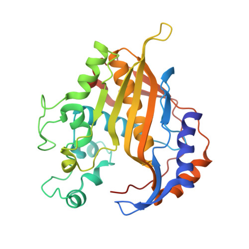
Approaches to solving the rigid receptor problem by identifying a minimal set of flexible residues during ligand docking.
Anderson, A.C., O'Neil, R.H., Surti, T.S., Stroud, R.M.(2001) Chem Biol 8: 445-457
- PubMed: 11358692
- DOI: https://doi.org/10.1016/s1074-5521(01)00023-0
- Primary Citation of Related Structures:
1F28 - PubMed Abstract:
Using fixed receptor sites derived from high-resolution crystal structures in structure-based drug design does not properly account for ligand-induced enzyme conformational change and imparts a bias into the discovery and design of novel ligands. We sought to facilitate the design of improved drug leads by defining residues most likely to change conformation, and then defining a minimal manifold of possible conformations of a target site for drug design based on a small number of identified flexible residues. The crystal structure of thymidylate synthase from an important pathogenic target Pneumocystis carinii (PcTS) bound to its substrate and the inhibitor, BW1843U89, is reported here and reveals a new conformation with respect to the structure of PcTS bound to substrate and the more conventional antifolate inhibitor, CB3717. We developed an algorithm for determining which residues provide 'soft spots' in the protein, regions where conformational adaptation suggests possible modifications for a drug lead that may yield higher affinity. Remodeling the active site of thymidylate synthase with new conformations for only three residues that were identified with this algorithm yields scores for ligands that are compatible with experimental kinetic data. Based on the examination of many protein/ligand complexes, we develop an algorithm (SOFTSPOTS) for identifying regions of a protein target that are more likely to accommodate plastically to regions of a drug molecule. Using these indicators we develop a second algorithm (PLASTIC) that provides a minimal manifold of possible conformations of a protein target for drug design, reducing the bias in structure-based drug design imparted by structures of enzymes co-crystallized with inhibitors.
Organizational Affiliation:
Department of Biochemistry and Biophysics, University of California at San Francisco, Box 0448, 94143-0448, USA. amy.c.anderson@dartmouth.edu


