1G8J

CRYSTAL STRUCTURE ANALYSIS OF ARSENITE OXIDASE FROM ALCALIGENES FAECALIS


Experimental Data Snapshot

  • Method: X-RAY DIFFRACTION
  • Resolution: 2.03 Å
  • R-Value Free: 0.223 
  • R-Value Work: 0.193 

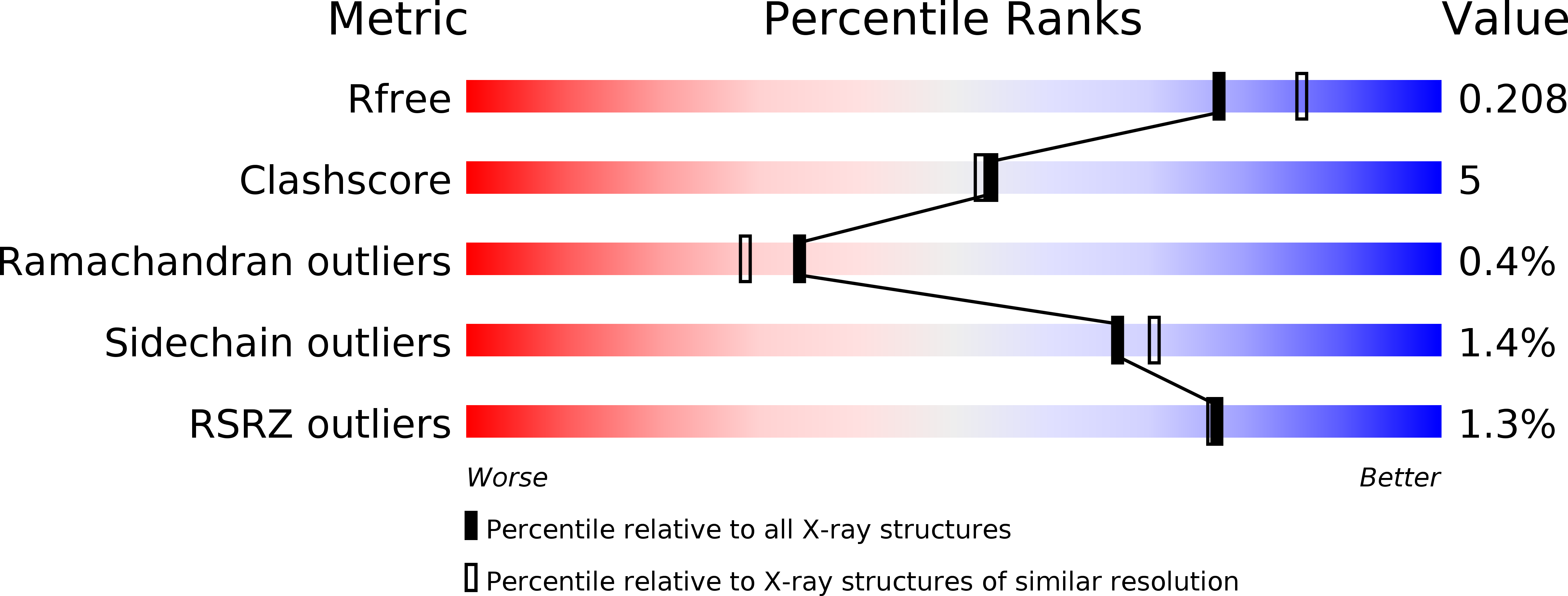
wwPDB Validation   3D Report Full Report


Ligand Structure Quality Assessment 


This is version 1.2 of the entry. See complete history


Literature

Crystal structure of the 100 kDa arsenite oxidase from Alcaligenes faecalis in two crystal forms at 1.64 A and 2.03 A.

Ellis, P.J.Conrads, T.Hille, R.Kuhn, P.

(2001) Structure 9: 125-132

  • DOI: https://doi.org/10.1016/s0969-2126(01)00566-4
  • Primary Citation of Related Structures:  
    1G8J, 1G8K

  • PubMed Abstract: 

    Arsenite oxidase from Alcaligenes faecalis NCIB 8687 is a molybdenum/iron protein involved in the detoxification of arsenic. It is induced by the presence of AsO(2-) (arsenite) and functions to oxidize As(III)O(2-), which binds to essential sulfhydryl groups of proteins and dithiols, to the relatively less toxic As(V)O(4)(3-) (arsenate) prior to methylation. Using a combination of multiple isomorphous replacement with anomalous scattering (MIRAS) and multiple-wavelength anomalous dispersion (MAD) methods, the crystal structure of arsenite oxidase was determined to 2.03 A in a P2(1) crystal form with two molecules in the asymmetric unit and to 1.64 A in a P1 crystal form with four molecules in the asymmetric unit. Arsenite oxidase consists of a large subunit of 825 residues and a small subunit of approximately 134 residues. The large subunit contains a Mo site, consisting of a Mo atom bound to two pterin cofactors, and a [3Fe-4S] cluster. The small subunit contains a Rieske-type [2Fe-2S] site. The large subunit of arsenite oxidase is similar to other members of the dimethylsulfoxide (DMSO) reductase family of molybdenum enzymes, particularly the dissimilatory periplasmic nitrate reductase from Desulfovibrio desulfuricans, but is unique in having no covalent bond between the polypeptide and the Mo atom. The small subunit has no counterpart among known Mo protein structures but is homologous to the Rieske [2Fe-2S] protein domain of the cytochrome bc(1) and cytochrome b(6)f complexes and to the Rieske domain of naphthalene 1,2-dioxygenase.


  • Organizational Affiliation

    Stanford Synchrotron Radiation Laboratory, Stanford University, 94309, Stanford, CA, USA.


Macromolecules
Find similar proteins by:  (by identity cutoff)  |  3D Structure
Entity ID: 1
MoleculeChains Sequence LengthOrganismDetailsImage
ARSENITE OXIDASE
A, C
825Alcaligenes faecalisMutation(s): 0 
EC: 1.20.9.1
UniProt
Find proteins for Q7SIF4 (Alcaligenes faecalis)
Explore Q7SIF4 
Go to UniProtKB:  Q7SIF4
Entity Groups  
Sequence Clusters30% Identity50% Identity70% Identity90% Identity95% Identity100% Identity
UniProt GroupQ7SIF4
Sequence Annotations
Expand
  • Reference Sequence
Find similar proteins by:  (by identity cutoff)  |  3D Structure
Entity ID: 2
MoleculeChains Sequence LengthOrganismDetailsImage
ARSENITE OXIDASE
B, D
133Alcaligenes faecalisMutation(s): 0 
EC: 1.20.9.1
UniProt
Find proteins for Q7SIF3 (Alcaligenes faecalis)
Explore Q7SIF3 
Go to UniProtKB:  Q7SIF3
Entity Groups  
Sequence Clusters30% Identity50% Identity70% Identity90% Identity95% Identity100% Identity
UniProt GroupQ7SIF3
Sequence Annotations
Expand
  • Reference Sequence
Small Molecules
Ligands 5 Unique
IDChains Name / Formula / InChI Key2D Diagram3D Interactions
MGD
Query on MGD

Download Ideal Coordinates CCD File 
E [auth A],
F [auth A],
K [auth C],
L [auth C]
2-AMINO-5,6-DIMERCAPTO-7-METHYL-3,7,8A,9-TETRAHYDRO-8-OXA-1,3,9,10-TETRAAZA-ANTHRACEN-4-ONE GUANOSINE DINUCLEOTIDE
C20 H26 N10 O13 P2 S2
VQAGYJCYOLHZDH-ILXWUORBSA-N
F3S
Query on F3S

Download Ideal Coordinates CCD File 
I [auth A],
O [auth C]
FE3-S4 CLUSTER
Fe3 S4
FCXHZBQOKRZXKS-UHFFFAOYSA-N
FES
Query on FES

Download Ideal Coordinates CCD File 
J [auth B],
P [auth D]
FE2/S2 (INORGANIC) CLUSTER
Fe2 S2
NIXDOXVAJZFRNF-UHFFFAOYSA-N
4MO
Query on 4MO

Download Ideal Coordinates CCD File 
H [auth A],
N [auth C]
MOLYBDENUM(IV) ION
Mo
ZIKKVZAYJJZBGE-UHFFFAOYSA-N
O
Query on O

Download Ideal Coordinates CCD File 
G [auth A],
M [auth C]
OXYGEN ATOM
O
XLYOFNOQVPJJNP-UHFFFAOYSA-N
Experimental Data & Validation

Experimental Data

  • Method: X-RAY DIFFRACTION
  • Resolution: 2.03 Å
  • R-Value Free: 0.223 
  • R-Value Work: 0.193 
  • Space Group: P 1 21 1
Unit Cell:
Length ( Å )Angle ( ˚ )
a = 96.71α = 90
b = 114.25β = 112.44
c = 108.98γ = 90
Software Package:
Software NamePurpose
MOSFLMdata reduction
TRUNCATEdata reduction
SHARPphasing
CNSrefinement
CCP4data scaling

Structure Validation

View Full Validation Report


Ligand Structure Quality Assessment 


Entry History 

Deposition Data

Revision History  (Full details and data files)

  • Version 1.0: 2000-12-13
    Type: Initial release
  • Version 1.1: 2008-04-27
    Changes: Version format compliance
  • Version 1.2: 2011-07-13
    Changes: Version format compliance