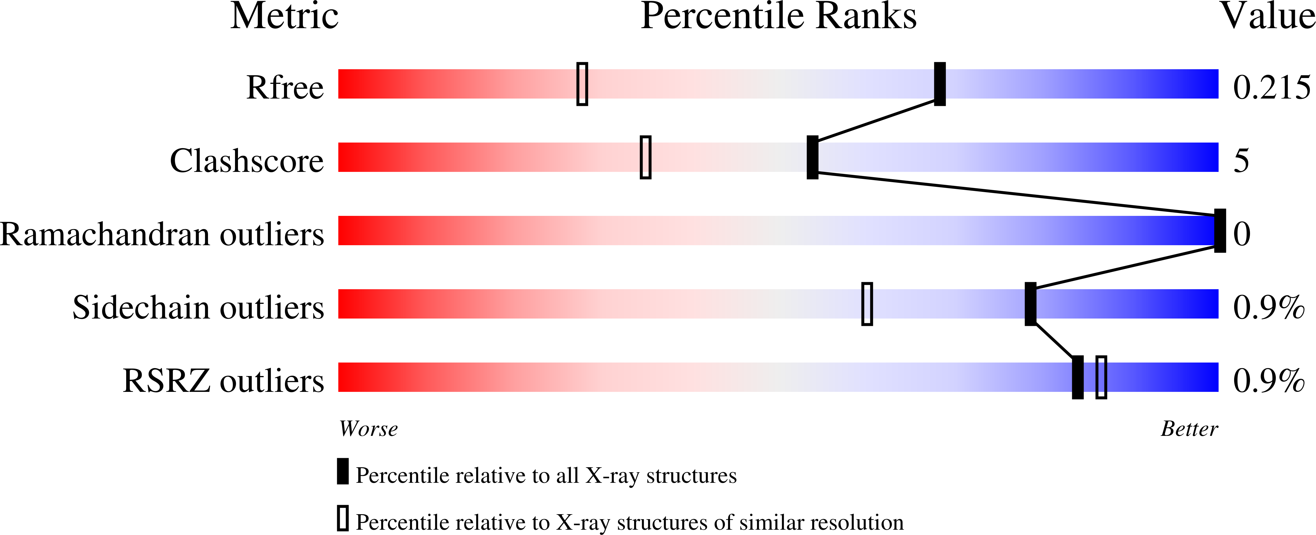
1H46

The catalytic module of Cel7D from Phanerochaete chrysosporium as a chiral selector: Structural studies of its complex with the b-blocker (R)-propranolol


Experimental Data Snapshot

  • Method: X-RAY DIFFRACTION
  • Resolution: 1.52 Å
  • R-Value Free: 0.219 
  • R-Value Work: 0.169 
  • R-Value Observed: 0.171 

Starting Model: experimental
View more details

wwPDB Validation   3D Report Full Report


Ligand Structure Quality Assessment 


This is version 2.2 of the entry. See complete history


Literature

The Catalytic Module of Cel7D from Phanerochaete Chrysosporium as a Chiral Selector: Structural Studies of its Complex with the Beta Blocker (R)-Propranolol

Munoz, I.G.Mowbray, S.L.Stahlberg, J.

(2003) Acta Crystallogr D Biol Crystallogr 59: 637

  • DOI: https://doi.org/10.1107/s0907444903001938
  • Primary Citation of Related Structures:  
    1H46

  • PubMed Abstract: 

    Previous investigations have shown that the major cellobiohydrolase of Phanerochaete chrysosporium, Cel7D (CBH 58), can be used to separate the enantiomers of a number of drugs, including adrenergic beta blockers such as propranolol. The structural basis of this enantioselectivity is explored here. A 1.5 A X-ray structure of the catalytic domain of Cel7D in complex with (R)-propranolol showed the ligand bound at the active site in glucosyl-binding subsites -1/+1. The catalytic residue Glu207 makes a strong charge-charge interaction with the secondary amine of (R)-propranolol; this is supported by a second interaction of the amine with the nearby Asp209. The aromatic naphthyl group stacks onto the indole ring of Trp373 (normally the glucosyl-binding platform of subsite +1). Other factors also contribute to good complementarity between the ligand and the substrate-binding cleft of the enzyme. Comparison with the previous structure of a related cellulase, Cel7A from Trichoderma reesei, in complex with (S)-propranolol strongly suggests that these enzymes will bind the (S)-enantiomer in a very similar manner, distinct from their mode of binding to (R)-propranolol. Tighter binding of both enzymes to the (S)-enantiomer is largely explained by two additional hydrogen-bonding interactions with its hydroxyl group. The distinct preference for the (R)-enantiomer is probably a consequence of structural differences near the naphthyl group of the ligand.


  • Organizational Affiliation

    Department of Molecular Biology, Swedish University of Agricultural Sciences, Uppsala.


Macromolecules
Find similar proteins by:  (by identity cutoff)  |  3D Structure
Entity ID: 1
MoleculeChains Sequence LengthOrganismDetailsImage
EXOGLUCANASE IA [auth X]431Phanerodontia chrysosporiumMutation(s): 0 
EC: 3.2.1.91 (PDB Primary Data), 3.2.1 (UniProt)
UniProt
Find proteins for Q7LHI2 (Phanerodontia chrysosporium)
Explore Q7LHI2 
Go to UniProtKB:  Q7LHI2
Entity Groups  
Sequence Clusters30% Identity50% Identity70% Identity90% Identity95% Identity100% Identity
UniProt GroupQ7LHI2
Glycosylation
Glycosylation Sites: 1
Sequence Annotations
Expand
  • Reference Sequence
Small Molecules
Ligands 2 Unique
IDChains Name / Formula / InChI Key2D Diagram3D Interactions
RNP
Query on RNP

Download Ideal Coordinates CCD File 
C [auth X](1E,2R)-1-(ISOPROPYLIMINO)-3-(1-NAPHTHYLOXY)PROPAN-2-OL
C16 H19 N O2
LLOTXRYLQVWOGC-AYCSXLNKSA-N
NAG
Query on NAG

Download Ideal Coordinates CCD File 
B [auth X]2-acetamido-2-deoxy-beta-D-glucopyranose
C8 H15 N O6
OVRNDRQMDRJTHS-FMDGEEDCSA-N
Modified Residues  1 Unique
IDChains TypeFormula2D DiagramParent
PCA
Query on PCA
A [auth X]L-PEPTIDE LINKINGC5 H7 N O3GLN
Binding Affinity Annotations 
IDSourceBinding Affinity
RNP PDBBind:  1H46 Ki: 2.70e+5 (nM) from 1 assay(s)
Experimental Data & Validation

Experimental Data

  • Method: X-RAY DIFFRACTION
  • Resolution: 1.52 Å
  • R-Value Free: 0.219 
  • R-Value Work: 0.169 
  • R-Value Observed: 0.171 
  • Space Group: C 1 2 1
Unit Cell:
Length ( Å )Angle ( ˚ )
a = 86.143α = 90
b = 46.557β = 102.99
c = 98.827γ = 90
Software Package:
Software NamePurpose
REFMACrefinement
MOSFLMdata reduction
SCALAdata scaling

Structure Validation

View Full Validation Report


Ligand Structure Quality Assessment 


Entry History 

Deposition Data

Revision History  (Full details and data files)

  • Version 1.0: 2003-04-03
    Type: Initial release
  • Version 1.1: 2011-05-08
    Changes: Version format compliance
  • Version 1.2: 2011-07-13
    Changes: Version format compliance
  • Version 1.3: 2019-05-29
    Changes: Data collection, Derived calculations, Experimental preparation, Other
  • Version 2.0: 2020-03-11
    Changes: Data collection, Other, Polymer sequence
  • Version 2.1: 2020-07-29
    Type: Remediation
    Reason: Carbohydrate remediation
    Changes: Data collection, Derived calculations, Structure summary
  • Version 2.2: 2023-12-13
    Changes: Data collection, Database references, Refinement description, Structure summary