1KZU

INTEGRAL MEMBRANE PERIPHERAL LIGHT HARVESTING COMPLEX FROM RHODOPSEUDOMONAS ACIDOPHILA STRAIN 10050


Experimental Data Snapshot

  • Method: X-RAY DIFFRACTION
  • Resolution: 2.50 Å
  • R-Value Free: 0.252 
  • R-Value Work: 0.227 
  • R-Value Observed: 0.227 

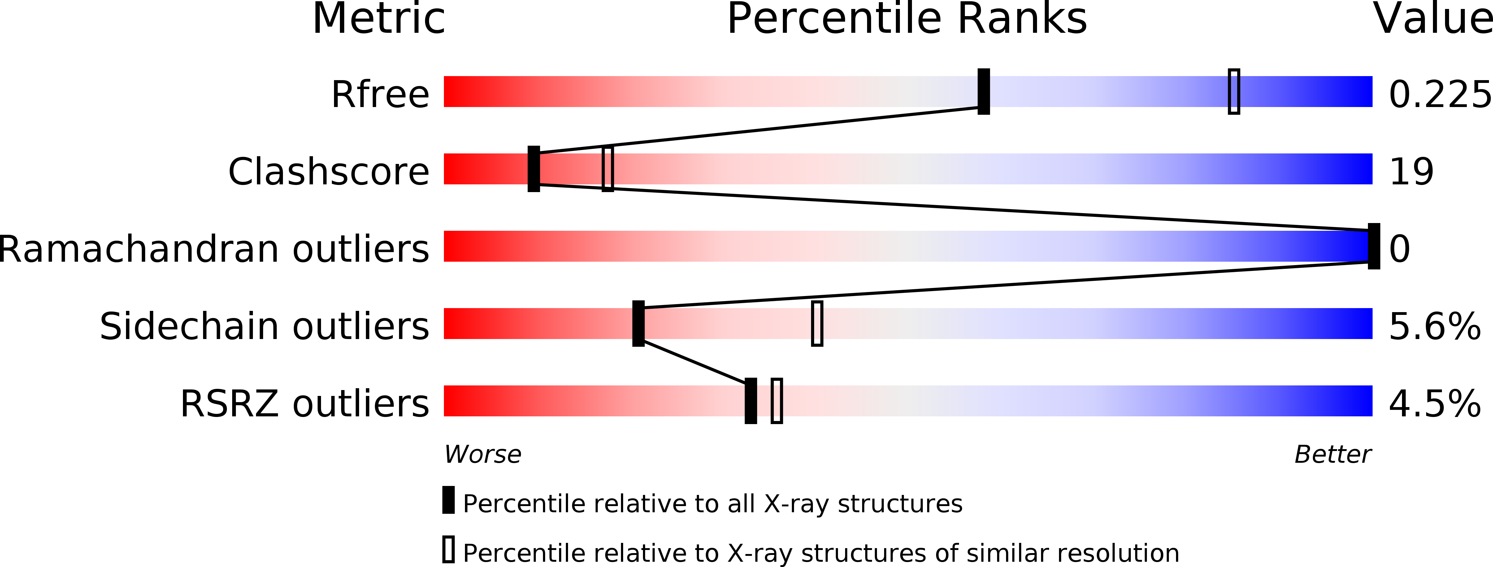
wwPDB Validation   3D Report Full Report


Ligand Structure Quality Assessment 


This is version 2.1 of the entry. See complete history


Literature

Apoprotein structure in the LH2 complex from Rhodopseudomonas acidophila strain 10050: modular assembly and protein pigment interactions.

Prince, S.M.Papiz, M.Z.Freer, A.A.McDermott, G.Hawthornthwaite-Lawless, A.M.Cogdell, R.J.Isaacs, N.W.

(1997) J Mol Biol 268: 412-423

  • DOI: https://doi.org/10.1006/jmbi.1997.0966
  • Primary Citation of Related Structures:  
    1KZU

  • PubMed Abstract: 

    The refined structure of the peripheral light-harvesting complex from Rhodopseudomonas acidophila strain 10050 reveals a membrane protein with protein-protein interactions in the trans-membrane region exclusively of a van der Waals nature. The dominant factors in the formation of the complex appear to be extramembranous hydrogen bonds (suggesting that each apoprotein must achieve a fold close to its final structure in order to oligomerize), protein-pigment and pigment-pigment interactions within the membrane-spanning region. The pigment molecules are known to play an important role in the formation of bacterial light-harvesters, and their extensive mediation of structural contacts within the membrane bears this out. Amino acid residues determining the secondary structure of the apoproteins influence the oligomeric state of the complex. The assembly of the pigment array is governed by the apoproteins of LH2. The particular environment of each of the pigment molecules is, however, influenced directly by few protein contacts. These contacts produce functional effects that are not attributable to a single cause, e.g. the arrangement of an overlapping cycle of chromophores not only provides energy delocalisation and storage properties, but also has consequences for oligomer size, pigment distortion modes and pigment chemical environment, all of which modify the precise function of the complex. The evaluation of site energies for the pigment array requires the consideration of a number of effects, including heterogeneous pigment distortions, charge distributions in the local environment and mechanical interactions.


  • Organizational Affiliation

    Department of Chemistry, University of Glasgow, UK.


Macromolecules
Find similar proteins by:  (by identity cutoff)  |  3D Structure
Entity ID: 1
MoleculeChains Sequence LengthOrganismDetailsImage
LIGHT HARVESTING PROTEIN B-800/850A,
C [auth D],
E [auth G]
53Rhodoblastus acidophilusMutation(s): 0 
Membrane Entity: Yes 
UniProt
Find proteins for P26789 (Rhodoblastus acidophilus)
Explore P26789 
Go to UniProtKB:  P26789
Entity Groups  
Sequence Clusters30% Identity50% Identity70% Identity90% Identity95% Identity100% Identity
UniProt GroupP26789
Sequence Annotations
Expand
  • Reference Sequence
Find similar proteins by:  (by identity cutoff)  |  3D Structure
Entity ID: 2
MoleculeChains Sequence LengthOrganismDetailsImage
LIGHT HARVESTING PROTEIN B-800/850B,
D [auth E],
F [auth H]
41Rhodoblastus acidophilusMutation(s): 0 
Membrane Entity: Yes 
UniProt
Find proteins for P26790 (Rhodoblastus acidophilus)
Explore P26790 
Go to UniProtKB:  P26790
Entity Groups  
Sequence Clusters30% Identity50% Identity70% Identity90% Identity95% Identity100% Identity
UniProt GroupP26790
Sequence Annotations
Expand
  • Reference Sequence
Small Molecules
Ligands 2 Unique
IDChains Name / Formula / InChI Key2D Diagram3D Interactions
BCL
Query on BCL

Download Ideal Coordinates CCD File 
H [auth A]
I [auth A]
K [auth B]
M [auth D]
N [auth D]
H [auth A],
I [auth A],
K [auth B],
M [auth D],
N [auth D],
P [auth E],
R [auth G],
S [auth G],
U [auth H]
BACTERIOCHLOROPHYLL A
C55 H74 Mg N4 O6
DSJXIQQMORJERS-AGGZHOMASA-M
RG1
Query on RG1

Download Ideal Coordinates CCD File 
G [auth A]
J [auth B]
L [auth D]
O [auth E]
Q [auth G]
G [auth A],
J [auth B],
L [auth D],
O [auth E],
Q [auth G],
T [auth H]
Rhodopin b-D-glucoside
C46 H66 O6
ISHBHDBCVQRMDY-GZIKAPSJSA-N
Modified Residues  1 Unique
IDChains TypeFormula2D DiagramParent
FME
Query on FME
A,
C [auth D],
E [auth G]
L-PEPTIDE LINKINGC6 H11 N O3 SMET
Experimental Data & Validation

Experimental Data

  • Method: X-RAY DIFFRACTION
  • Resolution: 2.50 Å
  • R-Value Free: 0.252 
  • R-Value Work: 0.227 
  • R-Value Observed: 0.227 
  • Space Group: H 3 2
Unit Cell:
Length ( Å )Angle ( ˚ )
a = 120.3α = 90
b = 120.3β = 90
c = 296.2γ = 120
Software Package:
Software NamePurpose
X-PLORmodel building
X-PLORrefinement
MOSFLMdata reduction
CCP4data scaling
ROTAVATAdata scaling
X-PLORphasing

Structure Validation

View Full Validation Report


Ligand Structure Quality Assessment 


Entry History 

Deposition Data

Revision History  (Full details and data files)

  • Version 1.0: 1997-04-01
    Type: Initial release
  • Version 1.1: 2008-03-24
    Changes: Version format compliance
  • Version 1.2: 2011-07-13
    Changes: Version format compliance
  • Version 1.3: 2016-03-30
    Changes: Non-polymer description
  • Version 2.0: 2024-06-05
    Changes: Advisory, Atomic model, Data collection, Database references, Derived calculations, Other, Refinement description
  • Version 2.1: 2024-11-06
    Changes: Advisory, Derived calculations, Structure summary