1LEE

CRYSTAL STRUCTURE OF PLASMEPSIN FROM P. FALCIPARUM IN COMPLEX WITH INHIBITOR RS367


Experimental Data Snapshot

  • Method: X-RAY DIFFRACTION
  • Resolution: 1.90 Å
  • R-Value Free: 0.286 
  • R-Value Work: 0.236 

wwPDB Validation   3D Report Full Report


This is version 1.4 of the entry. See complete history


Literature

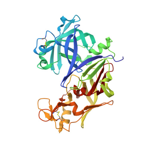
Structures of Ser205 mutant plasmepsin II from Plasmodium falciparum at 1.8 A in complex with the inhibitors rs367 and rs370.

Asojo, O.A.Afonina, E.Gulnik, S.V.Yu, B.Erickson, J.W.Randad, R.Medjahed, D.Silva, A.M.

(2002) Acta Crystallogr D Biol Crystallogr 58: 2001-2008

  • DOI: https://doi.org/10.1107/s0907444902014695
  • Primary Citation of Related Structures:  
    1LEE, 1LF2

  • PubMed Abstract: 

    Plasmepsin II is one of the four catalytically active plasmepsins found in the food vacuole of Plasmodium falciparum. These enzymes initiate hemoglobin degradation by cleavage at the alpha-chain between Phe33 and Leu34. The crystal structures of Ser205 mutant plasmepsin II from P. falciparum in complex with two inhibitors have been refined at a resolution of 1.8 A in the space group I222 and to R factors of 19.9 and 19.5%. Each crystal contains one monomer in the asymmetric unit. Both inhibitors have a Phe-Leu core and incorporate tetrahedral transition-state mimetic hydroxypropylamine. The inhibitor rs367 possesses a 2,6-dimethylphenyloxyacetyl group at the P2 position and 3-aminobenzamide at the P2' position, while rs370 has the same P2 group but 4-aminobenzamide in the P2' position. These complexes reveal key conserved hydrogen bonds between the inhibitor and the binding-cavity residues, notably with the flap residues Val78 and Ser79, the catalytic dyad Asp34 and Asp214 and the residues Ser218 and Gly36 that are in proximity to the catalytic dyad. The structures also show unexpected conformational variability of the binding cavity of plasmepsin II and may reflect the mode of binding of the hemoglobin alpha-chain for cleavage.


  • Organizational Affiliation

    Structure Biochemistry Program, National Cancer Institute/SAIC, Frederick, MD 21702, USA. asojo@ncifcrf.gov


Macromolecules
Find similar proteins by:  (by identity cutoff)  |  3D Structure
Entity ID: 1
MoleculeChains Sequence LengthOrganismDetailsImage
Plasmepsin 2331Plasmodium falciparumMutation(s): 1 
EC: 3.4.23.39
UniProt
Find proteins for P46925 (Plasmodium falciparum (isolate HB3))
Explore P46925 
Go to UniProtKB:  P46925
Entity Groups  
Sequence Clusters30% Identity50% Identity70% Identity90% Identity95% Identity100% Identity
UniProt GroupP46925
Sequence Annotations
Expand
  • Reference Sequence
Small Molecules
Ligands 1 Unique
IDChains Name / Formula / InChI Key2D Diagram3D Interactions
R36
Query on R36

Download Ideal Coordinates CCD File 
B [auth A]4-AMINO-N-{4-[2-(2,6-DIMETHYL-PHENOXY)-ACETYLAMINO]-3-HYDROXY-1-ISOBUTYL-5-PHENYL-PENTYL}-BENZAMIDE
C32 H41 N3 O4
WQUBEIMCFHCJCO-AWCRTANDSA-N
Binding Affinity Annotations 
IDSourceBinding Affinity
R36 PDBBind:  1LEE Ki: 18 (nM) from 1 assay(s)
Experimental Data & Validation

Experimental Data

  • Method: X-RAY DIFFRACTION
  • Resolution: 1.90 Å
  • R-Value Free: 0.286 
  • R-Value Work: 0.236 
  • Space Group: I 2 2 2
Unit Cell:
Length ( Å )Angle ( ˚ )
a = 75.89α = 90
b = 84.83β = 90
c = 123.08γ = 90
Software Package:
Software NamePurpose
AMoREphasing
CNSrefinement

Structure Validation

View Full Validation Report


Entry History 

Deposition Data

Revision History  (Full details and data files)

  • Version 1.0: 2002-10-09
    Type: Initial release
  • Version 1.1: 2008-04-28
    Changes: Version format compliance
  • Version 1.2: 2011-07-13
    Changes: Derived calculations, Version format compliance
  • Version 1.3: 2021-10-27
    Changes: Database references, Derived calculations
  • Version 1.4: 2023-08-16
    Changes: Data collection, Refinement description