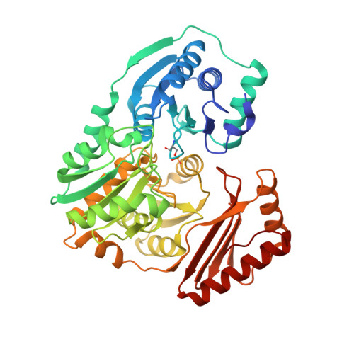
1P5G

Enzyme-ligand complex of P. aeruginosa PMM/PGM


Experimental Data Snapshot

  • Method: X-RAY DIFFRACTION
  • Resolution: 1.61 Å
  • R-Value Free: 0.187 
  • R-Value Work: 0.160 
  • R-Value Observed: 0.162 

Starting Model: experimental
View more details

wwPDB Validation   3D Report Full Report


Ligand Structure Quality Assessment 


This is version 1.6 of the entry. See complete history


Literature

Structural basis of diverse substrate recognition by the enzyme PMM/PGM from P. aeruginosa.

Regni, C.Naught, L.Tipton, P.A.Beamer, L.J.

(2004) Structure 12: 55-63

  • DOI: https://doi.org/10.1016/j.str.2003.11.015
  • Primary Citation of Related Structures:  
    1P5D, 1P5G, 1PCJ, 1PCM

  • PubMed Abstract: 

    Enzyme-substrate complexes of phosphomannomutase/phosphoglucomutase (PMM/PGM) reveal the structural basis of the enzyme's ability to use four different substrates in catalysis. High-resolution structures with glucose 1-phosphate, glucose 6-phosphate, mannose 1-phosphate, and mannose 6-phosphate show that the position of the phosphate group of each substrate is held constant by a conserved network of hydrogen bonds. This produces two distinct, and mutually exclusive, binding orientations for the sugar rings of the 1-phospho and 6-phospho sugars. Specific binding of both orientations is accomplished by key contacts with the O3 and O4 hydroxyls of the sugar, which must occupy equatorial positions. Dual recognition of glucose and mannose phosphosugars uses a combination of specific protein contacts and nonspecific solvent contacts. The ability of PMM/PGM to accommodate these four diverse substrates in a single active site is consistent with its highly reversible phosphoryl transfer reaction and allows it to function in multiple biosynthetic pathways in P. aeruginosa.


  • Organizational Affiliation

    Department of Biochemistry, 117 Schweitzer Hall, University of Missouri-Columbia, Columbia, MO 65211, USA.


Macromolecules
Find similar proteins by:  (by identity cutoff)  |  3D Structure
Entity ID: 1
MoleculeChains Sequence LengthOrganismDetailsImage
PhosphomannomutaseA [auth X]463Pseudomonas aeruginosaMutation(s): 0 
Gene Names: ALGC OR PA5322
EC: 5.4.2.8 (PDB Primary Data), 5.4.2.2 (UniProt)
UniProt
Find proteins for P26276 (Pseudomonas aeruginosa (strain ATCC 15692 / DSM 22644 / CIP 104116 / JCM 14847 / LMG 12228 / 1C / PRS 101 / PAO1))
Explore P26276 
Go to UniProtKB:  P26276
Entity Groups  
Sequence Clusters30% Identity50% Identity70% Identity90% Identity95% Identity100% Identity
UniProt GroupP26276
Sequence Annotations
Expand
  • Reference Sequence
Small Molecules
Modified Residues  1 Unique
IDChains TypeFormula2D DiagramParent
SEP
Query on SEP
A [auth X]L-PEPTIDE LINKINGC3 H8 N O6 PSER
Experimental Data & Validation

Experimental Data

  • Method: X-RAY DIFFRACTION
  • Resolution: 1.61 Å
  • R-Value Free: 0.187 
  • R-Value Work: 0.160 
  • R-Value Observed: 0.162 
  • Space Group: P 21 21 21
Unit Cell:
Length ( Å )Angle ( ˚ )
a = 70.591α = 90
b = 74.372β = 90
c = 85.994γ = 90
Software Package:
Software NamePurpose
REFMACrefinement
HKL-2000data reduction
SCALEPACKdata scaling
CNSphasing

Structure Validation

View Full Validation Report


Ligand Structure Quality Assessment 


Entry History 

Deposition Data

Revision History  (Full details and data files)

  • Version 1.0: 2004-01-20
    Type: Initial release
  • Version 1.1: 2008-04-29
    Changes: Version format compliance
  • Version 1.2: 2011-07-13
    Changes: Advisory, Version format compliance
  • Version 1.3: 2020-06-17
    Changes: Advisory, Database references, Derived calculations
  • Version 1.4: 2020-07-29
    Type: Remediation
    Reason: Carbohydrate remediation
    Changes: Data collection, Derived calculations, Structure summary
  • Version 1.5: 2023-08-16
    Changes: Data collection, Database references, Refinement description, Structure summary
  • Version 1.6: 2024-11-13
    Changes: Structure summary