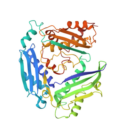
1P7L

S-Adenosylmethionine synthetase complexed with AMPPNP and Met.


Experimental Data Snapshot

  • Method: X-RAY DIFFRACTION
  • Resolution: 2.50 Å
  • R-Value Free: 0.242 
  • R-Value Work: 0.213 
  • R-Value Observed: 0.214 

wwPDB Validation   3D Report Full Report


This is version 1.4 of the entry. See complete history


Literature

Crystal structure of the s-adenosylmethionine synthetase ternary complex: a novel catalytic mechanism of s-adenosylmethionine synthesis from ATP and MET.

Komoto, J.Yamada, T.Takata, Y.Markham, G.D.Takusagawa, F.

(2004) Biochemistry 43: 1821-1831

  • DOI: https://doi.org/10.1021/bi035611t
  • Primary Citation of Related Structures:  
    1P7L, 1RG9

  • PubMed Abstract: 

    S-Adenosylmethionine synthetase (MAT) catalyzes formation of S-adenosylmethionine (SAM) from ATP and l-methionine (Met) and hydrolysis of tripolyphosphate to PP(i) and P(i). Escherichia coli MAT (eMAT) has been crystallized with the ATP analogue AMPPNP and Met, and the crystal structure has been determined at 2.5 A resolution. eMAT is a dimer of dimers and has a 222 symmetry. Each active site contains the products SAM and PPNP. A modeling study indicates that the substrates (AMPPNP and Met) can bind at the same sites as the products, and only a small conformation change of the ribose ring is needed for conversion of the substrates to the products. On the basis of the ternary complex structure and a modeling study, a novel catalytic mechanism of SAM formation is proposed. In the mechanism, neutral His14 acts as an acid to cleave the C5'-O5' bond of ATP while simultaneously a change in the ribose ring conformation from C4'-exo to C3'-endo occurs, and the S of Met makes a nucleophilic attack on the C5' to form SAM. All essential amino acid residues for substrate binding found in eMAT are conserved in the rat liver enzyme, indicating that the bacterial and mammalian enzymes have the same catalytic mechanism. However, a catalytic mechanism proposed recently by González et al. based on the structures of three ternary complexes of rat liver MAT [González, B., Pajares, M. A., Hermoso, J. A., Guillerm, D., Guillerm, G., and Sanz-Aparicio. J. (2003) J. Mol. Biol. 331, 407] is substantially different from our mechanism.


  • Organizational Affiliation

    Department of Molecular Biosciences, University of Kansas, 1200 Sunnyside Avenue, Lawrence, Kansas 66045-7534, USA.


Macromolecules
Find similar proteins by:  (by identity cutoff)  |  3D Structure
Entity ID: 1
MoleculeChains Sequence LengthOrganismDetailsImage
S-adenosylmethionine synthetase
A, B, C, D
383Escherichia coliMutation(s): 0 
Gene Names: METK OR METX OR B2942 OR C3528 OR Z4287 OR ECS3818 OR SF2933
EC: 2.5.1.6
UniProt
Find proteins for P0A817 (Escherichia coli (strain K12))
Explore P0A817 
Go to UniProtKB:  P0A817
Entity Groups  
Sequence Clusters30% Identity50% Identity70% Identity90% Identity95% Identity100% Identity
UniProt GroupP0A817
Sequence Annotations
Expand
  • Reference Sequence
Small Molecules
Ligands 6 Unique
IDChains Name / Formula / InChI Key2D Diagram3D Interactions
ANP
Query on ANP

Download Ideal Coordinates CCD File 
H [auth A],
N [auth B]
PHOSPHOAMINOPHOSPHONIC ACID-ADENYLATE ESTER
C10 H17 N6 O12 P3
PVKSNHVPLWYQGJ-KQYNXXCUSA-N
SAM
Query on SAM

Download Ideal Coordinates CCD File 
R [auth C],
S [auth C]
S-ADENOSYLMETHIONINE
C15 H22 N6 O5 S
MEFKEPWMEQBLKI-FCKMPRQPSA-N
PPK
Query on PPK

Download Ideal Coordinates CCD File 
T [auth C],
X [auth D]
(DIPHOSPHONO)AMINOPHOSPHONIC ACID
H6 N O9 P3
PELPUMGXMYVGSQ-UHFFFAOYSA-N
MET
Query on MET

Download Ideal Coordinates CCD File 
I [auth A],
M [auth B]
METHIONINE
C5 H11 N O2 S
FFEARJCKVFRZRR-BYPYZUCNSA-N
K
Query on K

Download Ideal Coordinates CCD File 
E [auth A],
J [auth B],
O [auth C],
V [auth D]
POTASSIUM ION
K
NPYPAHLBTDXSSS-UHFFFAOYSA-N
MG
Query on MG

Download Ideal Coordinates CCD File 
F [auth A]
G [auth A]
K [auth B]
L [auth B]
P [auth C]
F [auth A],
G [auth A],
K [auth B],
L [auth B],
P [auth C],
Q [auth C],
U [auth D],
W [auth D]
MAGNESIUM ION
Mg
JLVVSXFLKOJNIY-UHFFFAOYSA-N
Experimental Data & Validation

Experimental Data

  • Method: X-RAY DIFFRACTION
  • Resolution: 2.50 Å
  • R-Value Free: 0.242 
  • R-Value Work: 0.213 
  • R-Value Observed: 0.214 
  • Space Group: P 21 21 21
Unit Cell:
Length ( Å )Angle ( ˚ )
a = 225.82α = 90
b = 69.13β = 90
c = 118.23γ = 90
Software Package:
Software NamePurpose
SCALEPACKdata scaling
X-PLORmodel building
X-PLORrefinement
X-PLORphasing

Structure Validation

View Full Validation Report


Entry History 

Deposition Data

Revision History  (Full details and data files)

  • Version 1.0: 2004-03-02
    Type: Initial release
  • Version 1.1: 2008-04-29
    Changes: Version format compliance
  • Version 1.2: 2011-07-13
    Changes: Version format compliance
  • Version 1.3: 2017-10-11
    Changes: Refinement description
  • Version 1.4: 2024-02-14
    Changes: Advisory, Data collection, Database references, Derived calculations