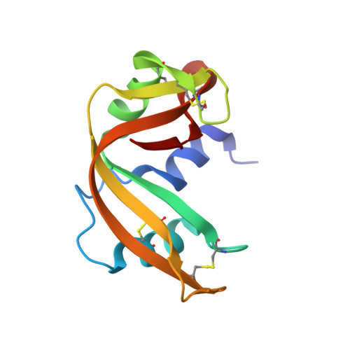
The structures of RNase A complexed with 3'-CMP and d(CpA): active site conformation and conserved water molecules.
Zegers, I., Maes, D., Dao-Thi, M.H., Poortmans, F., Palmer, R., Wyns, L.(1994) Protein Sci 3: 2322-2339
- PubMed: 7756988
- DOI: https://doi.org/10.1002/pro.5560031217
- Primary Citation of Related Structures:
1RPF, 1RPG, 1RPH - PubMed Abstract:
The interactions of RNase A with cytidine 3'-monophosphate (3'-CMP) and deoxycytidyl-3',5'-deoxyadenosine (d(CpA)) were analyzed by X-ray crystallography. The 3'-CMP complex and the native structure were determined from trigonal crystals, and the d(CpA) complex from monoclinic crystals. The differences between the overall structures are concentrated in loop regions and are relatively small. The protein-inhibitor contacts are interpreted in terms of the catalytic mechanism. The general base His 12 interacts with the 2' oxygen, as does the electrostatic catalyst Lys 41. The general acid His 119 has 2 conformations (A and B) in the native structure and is found in, respectively, the A and the B conformation in the d(CpA) and the 3'-CMP complex. From the present structures and from a comparison with RNase T1, we propose that His 119 is active in the A conformation. The structure of the d(CpA) complex permits a detailed analysis of the downstream binding site, which includes His 119 and Asn 71. The comparison of the present RNase A structures with an inhibitor complex of RNase T1 shows that there are important similarities in the active sites of these 2 enzymes, despite the absence of any sequence homology. The water molecules were analyzed in order to identify conserved water sites. Seventeen water sites were found to be conserved in RNase A structures from 5 different space groups. It is proposed that 7 of those water molecules play a role in the binding of the N-terminal helix to the rest of the protein and in the stabilization of the active site.
Organizational Affiliation:
Institute of Molecular Biology, Vrije Universiteit Brussel, St. Genesius Rode, Belgium.


