1RZT

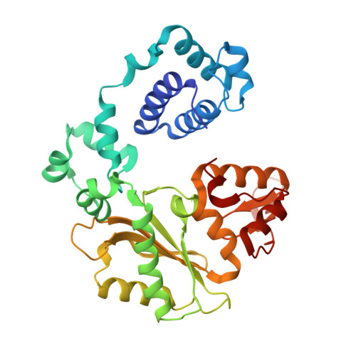
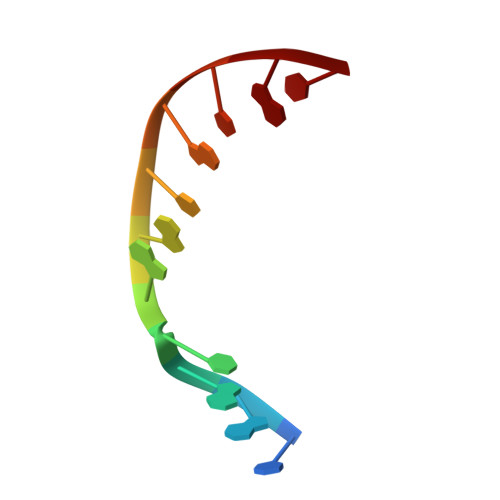
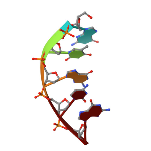
Crystal structure of DNA polymerase lambda complexed with a two nucleotide gap DNA molecule


Experimental Data Snapshot

  • Method: X-RAY DIFFRACTION
  • Resolution: 2.10 Å
  • R-Value Free: 0.260 
  • R-Value Work: 0.227 
  • R-Value Observed: 0.227 

Starting Model: experimental
View more details

wwPDB Validation   3D Report Full Report


This is version 1.5 of the entry. See complete history


Literature

A structural solution for the DNA polymerase lambda-dependent repair of DNA gaps with minimal homology.

Garcia-Diaz, M.Bebenek, K.Krahn, J.M.Blanco, L.Kunkel, T.A.Pedersen, L.C.

(2004) Mol Cell 13: 561-572

  • DOI: https://doi.org/10.1016/s1097-2765(04)00061-9
  • Primary Citation of Related Structures:  
    1RZT

  • PubMed Abstract: 

    Human DNA polymerase lambda (Pol lambda) is a family X member with low frameshift fidelity that has been suggested to perform gap-filling DNA synthesis during base excision repair and during repair of broken ends with limited homology. Here, we present a 2.1 A crystal structure of the catalytic core of Pol lambda in complex with DNA containing a two nucleotide gap. Pol lambda makes limited contacts with the template strand at the polymerase active site, and superimposition with Pol beta in a ternary complex suggests a shift in the position of the DNA at the active site that is reminiscent of a deletion intermediate. Surprisingly, Pol lambda can adopt a closed conformation, even in the absence of dNTP binding. These observations have implications for the catalytic mechanism and putative DNA repair functions of Pol lambda.


  • Organizational Affiliation

    Laboratory of Molecular Genetics, National Institute of Environmental Health Sciences, Research Triangle Park, NC 27709, USA.


Macromolecules

Find similar proteins by:  (by identity cutoff)  |  3D Structure
Entity ID: 4
MoleculeChains Sequence LengthOrganismDetailsImage
DNA polymerase lambdaM [auth A],
N [auth E],
O [auth I],
P [auth M]
331Homo sapiensMutation(s): 0 
Gene Names: POLL
EC: 2.7.7.7 (PDB Primary Data), 4.2.99 (UniProt)
UniProt & NIH Common Fund Data Resources
Find proteins for Q9UGP5 (Homo sapiens)
Explore Q9UGP5 
Go to UniProtKB:  Q9UGP5
PHAROS:  Q9UGP5
GTEx:  ENSG00000166169 
Entity Groups  
Sequence Clusters30% Identity50% Identity70% Identity90% Identity95% Identity100% Identity
UniProt GroupQ9UGP5
Sequence Annotations
Expand
  • Reference Sequence

Find similar nucleic acids by:  Sequence   |   3D Structure  

Entity ID: 1
MoleculeChains LengthOrganismImage
5'-D(*CP*GP*GP*CP*AP*AP*CP*GP*CP*AP*C)-3'A [auth B],
D [auth F],
G [auth J],
J [auth N]
11N/A
Sequence Annotations
Expand
  • Reference Sequence

Find similar nucleic acids by:  Sequence   |   3D Structure  

Entity ID: 2
MoleculeChains LengthOrganismImage
5'-D(*GP*TP*GP*CP*G)-3'B [auth C],
E [auth G],
H [auth K],
K [auth O]
5N/A
Sequence Annotations
Expand
  • Reference Sequence

Find similar nucleic acids by:  Sequence   |   3D Structure  

Entity ID: 3
MoleculeChains LengthOrganismImage
5'-D(P*GP*CP*CP*G)-3'C [auth D],
F [auth H],
I [auth L],
L [auth P]
4N/A
Sequence Annotations
Expand
  • Reference Sequence
Small Molecules
Ligands 2 Unique
IDChains Name / Formula / InChI Key2D Diagram3D Interactions
EDO
Query on EDO

Download Ideal Coordinates CCD File 
AA [auth M]1,2-ETHANEDIOL
C2 H6 O2
LYCAIKOWRPUZTN-UHFFFAOYSA-N
NA
Query on NA

Download Ideal Coordinates CCD File 
Q [auth B]
R [auth F]
S [auth J]
T [auth J]
U [auth A]
Q [auth B],
R [auth F],
S [auth J],
T [auth J],
U [auth A],
V [auth A],
W [auth E],
X [auth I],
Y [auth M],
Z [auth M]
SODIUM ION
Na
FKNQFGJONOIPTF-UHFFFAOYSA-N
Experimental Data & Validation

Experimental Data

  • Method: X-RAY DIFFRACTION
  • Resolution: 2.10 Å
  • R-Value Free: 0.260 
  • R-Value Work: 0.227 
  • R-Value Observed: 0.227 
  • Space Group: P 21 21 2
Unit Cell:
Length ( Å )Angle ( ˚ )
a = 191.321α = 90
b = 99.034β = 90
c = 104.838γ = 90
Software Package:
Software NamePurpose
CNSrefinement
SCALEPACKdata scaling
MOLREPphasing

Structure Validation

View Full Validation Report


Entry History 

Deposition Data

Revision History  (Full details and data files)

  • Version 1.0: 2004-03-02
    Type: Initial release
  • Version 1.1: 2008-04-29
    Changes: Version format compliance
  • Version 1.2: 2011-07-13
    Changes: Version format compliance
  • Version 1.3: 2017-10-11
    Changes: Refinement description
  • Version 1.4: 2024-04-03
    Changes: Data collection, Database references, Derived calculations, Refinement description
  • Version 1.5: 2024-10-09
    Changes: Structure summary