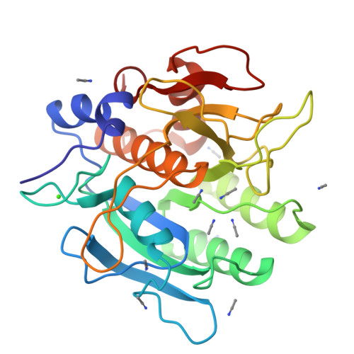
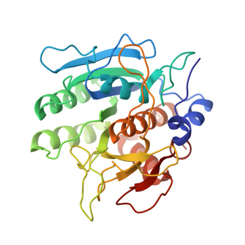
1SCB

ENZYME CRYSTAL STRUCTURE IN A NEAT ORGANIC SOLVENT


Experimental Data Snapshot

  • Method: X-RAY DIFFRACTION
  • Resolution: 2.30 Å
  • R-Value Observed: 0.184 

wwPDB Validation   3D Report Full Report


This is version 1.4 of the entry. See complete history


Literature

Enzyme crystal structure in a neat organic solvent.

Fitzpatrick, P.A.Steinmetz, A.C.Ringe, D.Klibanov, A.M.

(1993) Proc Natl Acad Sci U S A 90: 8653-8657

  • DOI: https://doi.org/10.1073/pnas.90.18.8653
  • Primary Citation of Related Structures:  
    1SCA, 1SCB

  • PubMed Abstract: 

    The crystal structure of the serine protease subtilisin Carlsberg in anhydrous acetonitrile was determined at 2.3 A resolution. It was found to be essentially identical to the three-dimensional structure of the enzyme in water; the differences observed were smaller than those between two independently determined structures in aqueous solution. The hydrogen bond system of the catalytic triad is intact in acetonitrile. The majority (99 of 119) of enzyme-bound, structural water molecules have such a great affinity to subtilisin that they are not displaced even in anhydrous acetonitrile. Of the 12 enzyme-bound acetonitrile molecules, 4 displace water molecules and 8 bind where no water had been observed before. One-third of all subtilisin-bound acetonitrile molecules reside in the active center, occupying the same region (P1, P2, and P3 binding sites) as the specific protein inhibitor eglin c.


  • Organizational Affiliation

    Department of Chemistry, Massachusetts Institute of Technology, Cambridge 02139.


Macromolecules
Find similar proteins by:  (by identity cutoff)  |  3D Structure
Entity ID: 1
MoleculeChains Sequence LengthOrganismDetailsImage
SUBTILISIN CARLSBERG274Bacillus licheniformisMutation(s): 0 
EC: 3.4.21.62
UniProt
Find proteins for P00780 (Bacillus licheniformis)
Explore P00780 
Go to UniProtKB:  P00780
Entity Groups  
Sequence Clusters30% Identity50% Identity70% Identity90% Identity95% Identity100% Identity
UniProt GroupP00780
Sequence Annotations
Expand
  • Reference Sequence
Small Molecules
Ligands 2 Unique
IDChains Name / Formula / InChI Key2D Diagram3D Interactions
CCN
Query on CCN

Download Ideal Coordinates CCD File 
C [auth A]
D [auth A]
E [auth A]
F [auth A]
G [auth A]
C [auth A],
D [auth A],
E [auth A],
F [auth A],
G [auth A],
H [auth A],
I [auth A],
J [auth A],
K [auth A],
L [auth A],
M [auth A],
N [auth A]
ACETONITRILE
C2 H3 N
WEVYAHXRMPXWCK-UHFFFAOYSA-N
CA
Query on CA

Download Ideal Coordinates CCD File 
B [auth A]CALCIUM ION
Ca
BHPQYMZQTOCNFJ-UHFFFAOYSA-N
Experimental Data & Validation

Experimental Data

  • Method: X-RAY DIFFRACTION
  • Resolution: 2.30 Å
  • R-Value Observed: 0.184 
  • Space Group: P 21 21 21
Unit Cell:
Length ( Å )Angle ( ˚ )
a = 77.1α = 90
b = 55.4β = 90
c = 53.6γ = 90
Software Package:
Software NamePurpose
TNTrefinement

Structure Validation

View Full Validation Report


Entry History 

Deposition Data

Revision History  (Full details and data files)

  • Version 1.0: 1994-01-31
    Type: Initial release
  • Version 1.1: 2008-03-03
    Changes: Version format compliance
  • Version 1.2: 2011-07-13
    Changes: Version format compliance
  • Version 1.3: 2017-11-29
    Changes: Derived calculations, Other
  • Version 1.4: 2024-02-14
    Changes: Data collection, Database references, Derived calculations