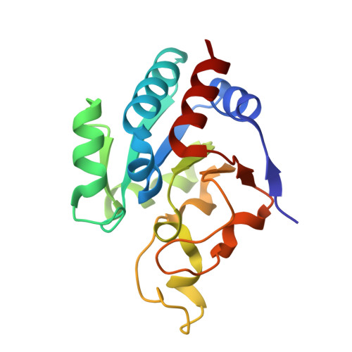
1XLT

Crystal structure of Transhydrogenase [(domain I)2:domain III] heterotrimer complex


Experimental Data Snapshot

  • Method: X-RAY DIFFRACTION
  • Resolution: 3.10 Å
  • R-Value Free: 0.310 
  • R-Value Work: 0.232 

wwPDB Validation   3D Report Full Report


Ligand Structure Quality Assessment 


This is version 2.1 of the entry. See complete history


Literature

Conformational Diversity in NAD(H) and Transhydrogenase Nicotinamide Nucleotide Binding Domains

Sundaresan, V.Chartron, J.Yamaguchi, M.Stout, C.D.

(2005) J Mol Biol 346: 617-629

  • DOI: https://doi.org/10.1016/j.jmb.2004.11.070
  • Primary Citation of Related Structures:  
    1XLT

  • PubMed Abstract: 

    Transhydrogenase (TH) couples direct and stereospecific hydride transfer between NAD(H) and NADP(H), bound within soluble domains I and III, respectively, to proton translocation across membrane bound domain II. The cocrystal structure of Rhodospirillum rubrum TH domains I and III has been determined in the presence of limiting NADH, under conditions in which the subunits reach equilibrium during crystallization. The crystals contain three heterotrimeric complexes, dI(2)dIII, in the asymmetric unit. Multiple conformations of loops and side-chains, and NAD(H) cofactors, are observed in domain I pertaining to substrate/product exchange, and highlighting electrostatic interactions during the hydride transfer. Two interacting NAD(H)-NADPH pairs are observed where alternate conformations of the NAD(H) phosphodiester and conserved arginine side-chains are correlated. In addition, the stereochemistry of one NAD(H)-NADPH pair approaches that expected for nicotinamide hydride transfer reactions. The cocrystal structure exhibits non-crystallographic symmetry that implies another orientation for domain III, which could occur in dimeric TH. Superposition of the "closed" form of domain III (PDB 1PNO, chain A) onto the dI(2)dIII complex reveals a severe steric conflict of highly conserved loops in domains I and III. This overlap, and the overlap with a 2-fold related domain III, suggests that motions of loop D within domain III and of the entire domain are correlated during turnover. The results support the concept that proton pumping in TH is driven by the difference in binding affinity for oxidized and reduced nicotinamide cofactors, and in the absence of a difference in redox potential, must occur through conformational effects.


  • Organizational Affiliation

    Department of Molecular Biology, The Scripps Research Institute, 10550 N. Torrey Pines Road, La Jolla, CA 92037, USA.


Macromolecules
Find similar proteins by:  (by identity cutoff)  |  3D Structure
Entity ID: 1
MoleculeChains Sequence LengthOrganismDetailsImage
NAD(P) transhydrogenase subunit alpha part 1
A, B, D, E, G
A, B, D, E, G, H
384Rhodospirillum rubrumMutation(s): 0 
Gene Names: pntAAnntA1
EC: 1.6.1.2 (PDB Primary Data), 7.1.1.1 (UniProt)
UniProt
Find proteins for Q2RSB2 (Rhodospirillum rubrum (strain ATCC 11170 / ATH 1.1.1 / DSM 467 / LMG 4362 / NCIMB 8255 / S1))
Explore Q2RSB2 
Go to UniProtKB:  Q2RSB2
Entity Groups  
Sequence Clusters30% Identity50% Identity70% Identity90% Identity95% Identity100% Identity
UniProt GroupQ2RSB2
Sequence Annotations
Expand
  • Reference Sequence
Find similar proteins by:  (by identity cutoff)  |  3D Structure
Entity ID: 2
MoleculeChains Sequence LengthOrganismDetailsImage
NAD(P) transhydrogenase subunit beta
C, F, I
174Rhodospirillum rubrumMutation(s): 0 
Gene Names: pntBnntB
EC: 1.6.1.2 (PDB Primary Data), 7.1.1.1 (UniProt)
UniProt
Find proteins for Q2RSB4 (Rhodospirillum rubrum (strain ATCC 11170 / ATH 1.1.1 / DSM 467 / LMG 4362 / NCIMB 8255 / S1))
Explore Q2RSB4 
Go to UniProtKB:  Q2RSB4
Entity Groups  
Sequence Clusters30% Identity50% Identity70% Identity90% Identity95% Identity100% Identity
UniProt GroupQ2RSB4
Sequence Annotations
Expand
  • Reference Sequence
Oligosaccharides

Help

Entity ID: 3
MoleculeChains Length2D Diagram Glycosylation3D Interactions
beta-D-fructofuranose-(2-1)-alpha-D-glucopyranose
J, K, L, M, N
J, K, L, M, N, O
2N/A
Glycosylation Resources
GlyTouCan:  G05551OP
GlyCosmos:  G05551OP
Small Molecules
Ligands 3 Unique
IDChains Name / Formula / InChI Key2D Diagram3D Interactions
NDP
Query on NDP

Download Ideal Coordinates CCD File 
S [auth C],
V [auth F],
Z [auth I]
NADPH DIHYDRO-NICOTINAMIDE-ADENINE-DINUCLEOTIDE PHOSPHATE
C21 H30 N7 O17 P3
ACFIXJIJDZMPPO-NNYOXOHSSA-N
NAD
Query on NAD

Download Ideal Coordinates CCD File 
P [auth A],
R [auth B],
T [auth D],
W [auth G],
Y [auth H]
NICOTINAMIDE-ADENINE-DINUCLEOTIDE
C21 H27 N7 O14 P2
BAWFJGJZGIEFAR-NNYOXOHSSA-N
NA
Query on NA

Download Ideal Coordinates CCD File 
Q [auth A],
U [auth D],
X [auth G]
SODIUM ION
Na
FKNQFGJONOIPTF-UHFFFAOYSA-N
Experimental Data & Validation

Experimental Data

  • Method: X-RAY DIFFRACTION
  • Resolution: 3.10 Å
  • R-Value Free: 0.310 
  • R-Value Work: 0.232 
  • Space Group: P 21 21 21
Unit Cell:
Length ( Å )Angle ( ˚ )
a = 95.372α = 90
b = 171.082β = 90
c = 203.181γ = 90
Software Package:
Software NamePurpose
CNSrefinement
MOSFLMdata reduction
CCP4data scaling
BEASTphasing

Structure Validation

View Full Validation Report


Ligand Structure Quality Assessment 


Entry History 

Deposition Data

Revision History  (Full details and data files)

  • Version 1.0: 2005-04-05
    Type: Initial release
  • Version 1.1: 2008-04-30
    Changes: Version format compliance
  • Version 1.2: 2011-07-13
    Changes: Version format compliance
  • Version 2.0: 2020-07-29
    Type: Remediation
    Reason: Carbohydrate remediation
    Changes: Advisory, Atomic model, Data collection, Database references, Derived calculations, Non-polymer description, Structure summary
  • Version 2.1: 2024-02-14
    Changes: Data collection, Database references, Structure summary