Unusual Entropy Driven Affinity of Chromobacterium Violaceum Lectin Cv-Iil Towards Fucose and Mannose
Pokorna, M., Cioci, G., Perret, S., Rebuffet, E., Kostlanova, N., Adam, J., Gilboa-Garber, N., Mitchell, E.P., Imberty, A., Wimmerova, M.(2006) Biochemistry 45: 7501
- PubMed: 16768446
- DOI: https://doi.org/10.1021/bi060214e
- Primary Citation of Related Structures:
2BOI, 2BV4 - PubMed Abstract:
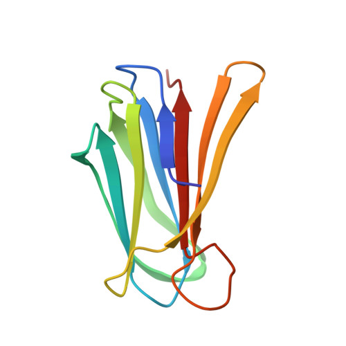
The purple pigmented bacterium Chromobacterium violaceum is a dominant component of tropical soil microbiota that can cause rare but fatal septicaemia in humans. Its sequenced genome provides insight into the abundant potential of this organism for biotechnological and pharmaceutical applications and allowed an ORF encoding a protein that is 60% identical to the fucose binding lectin (PA-IIL) from Pseudomonas aeruginosa and the mannose binding lectin (RS-IIL) from Ralstonia solanacearum to be identified. The lectin, CV-IIL, has recently been purified from C. violaceum [Zinger-Yosovich, K., Sudakevitz, D., Imberty, A., Garber, N. C., and Gilboa-Garber, N. (2006) Microbiology 152, 457-463] and has been confirmed to be a tetramer with subunit size of 11.86 kDa and a binding preference for fucose. We describe here the cloning of CV-IIL and its expression as a recombinant protein. A complete structure-function characterization has been made in an effort to analyze the specificity and affinity of CV-IIL for fucose and mannose. Crystal structures of CV-IIL complexes with monosaccharides have yielded the molecular basis of the specificity. Each monomer contains two close calcium cations that mediate the binding of the monosaccharides, which occurs in different orientations for fucose and mannose. The thermodynamics of binding has been analyzed by titration microcalorimetry, giving dissociation constants of 1.7 and 19 microM for alpha-methyl fucoside and alpha-methyl mannoside, respectively. Further analysis demonstrated a strongly favorable entropy term that is unusual in carbohydrate binding. A comparison with both PA-IIL and RS-IIL, which have binding preferences for fucose and mannose, respectively, yielded insights into the monosaccharide specificity of this important class of soluble bacterial lectins.
Organizational Affiliation:
National Centre for Biomolecular Research and Department of Biochemistry, Faculty of Science, Masaryk University, Kotlarska 2, 611 37 Brno, Czech Republic.


