2CAN | pdb_00002can

HUMAN ORNITHINE AMINOTRANSFERASE COMPLEXED WITH L-CANALINE


Experimental Data Snapshot

  • Method: X-RAY DIFFRACTION
  • Resolution: 2.30 Å
  • R-Value Free: 
    0.238 (Depositor) 
  • R-Value Work: 
    0.212 (Depositor), 0.220 (DCC) 
  • R-Value Observed: 
    0.212 (Depositor) 

Starting Model: experimental
View more details

wwPDB Validation   3D Report Full Report


Ligand Structure Quality Assessment 

Created with Raphaël 2.3.0Worse 01 BetterLigand structure goodness of fit to experimental dataBest fitted PLPClick on this verticalbar to view details

This is version 1.4 of the entry. See complete history


Literature

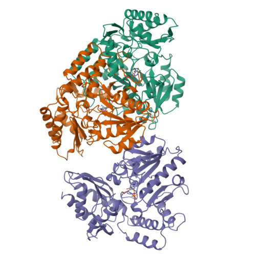
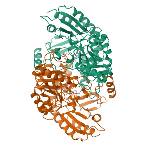
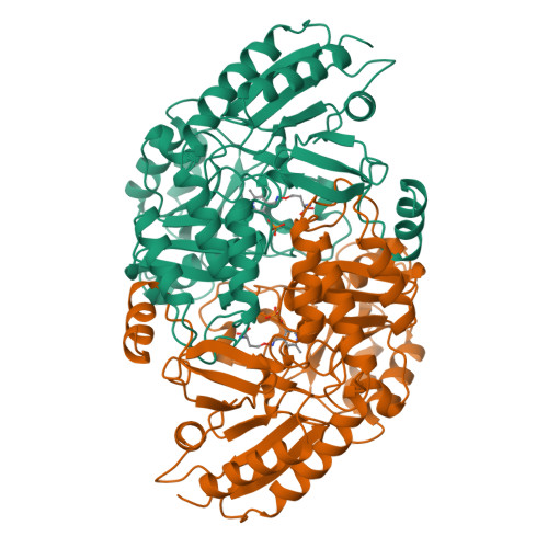
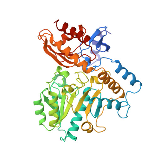
Human ornithine aminotransferase complexed with L-canaline and gabaculine: structural basis for substrate recognition.

Shah, S.A.Shen, B.W.Brunger, A.T.

(1997) Structure 5: 1067-1075

  • DOI: https://doi.org/10.1016/s0969-2126(97)00258-x
  • Primary Citation of Related Structures:  
    1GBN, 2CAN

  • PubMed Abstract: 

    Ornithine aminotransferase (OAT) is a 45 kDa pyridoxal-5'-phosphate (PLP)-dependent enzyme that catalyzes the conversion of L-ornithine and 2-oxoglutarate to glutamate-delta-semialdehyde and glutamic acid, respectively. In humans, loss of OAT function causes an accumulation of ornithine that results in gyrate atrophy of the choroid and retina, a disease that progressively leads to blindness. In an effort to learn more about the structural basis of this enzyme's function, we have determined the X-ray structures of OAT in complex with two enzyme-activated suicide substrates: L-canaline, an ornithine analog, and gabaculine, an irreversible inhibitor of several related aminotransferases. The structures of human OAT bound to the inhibitors gabaculine and L-canaline were solved to 2.3 A at 110K by difference Fourier techniques. Both inhibitors coordinate similarly in the active site, binding covalently to the PLP cofactor and causing a 20 degrees rotation in the cofactor tilt relative to the ligand-free form. Aromatic-aromatic interactions occur between the bound gabaculine molecule and active-site residues Tyr85 and Phe177, whereas Tyr55 and Arg180 provide specific contacts to the alpha-amino and carboxyl groups of L-canaline. The OAT-L-canaline complex structure implicates Tyr55 and Arg180 as the residues involved in coordinating with the natural substrate ornithine during normal enzyme turnover. This correlates well with two enzyme-inactivating point mutations associated with gyrate atrophy, Tyr55-->His and Arg180-->Thr. The OAT-gabaculine complex provides the first structural evidence that the potency of the inhibitor is due to energetically favourable aromatic interactions with residues in the active site. This aromatic-binding mode may be relevant to structure-based drug design efforts against other omega-aminotransferase targets, such as GABA aminotransferase.


  • Organizational Affiliation

    Department of Molecular Biophysics and Biochemistry, Yale University, New Haven, CT 06520, USA.


Macromolecules
Find similar proteins by:  (by identity cutoff)  |  3D Structure
Entity ID: 1
MoleculeChains Sequence LengthOrganismDetailsImage
ORNITHINE AMINOTRANSFERASE
A, B, C
402Homo sapiensMutation(s): 0 
EC: 2.6.1.13
UniProt & NIH Common Fund Data Resources
Find proteins for P04181 (Homo sapiens)
Explore P04181 
Go to UniProtKB:  P04181
PHAROS:  P04181
GTEx:  ENSG00000065154 
Entity Groups  
Sequence Clusters30% Identity50% Identity70% Identity90% Identity95% Identity100% Identity
UniProt GroupP04181
Sequence Annotations
Expand
  • Reference Sequence
Experimental Data & Validation

Experimental Data

  • Method: X-RAY DIFFRACTION
  • Resolution: 2.30 Å
  • R-Value Free:  0.238 (Depositor) 
  • R-Value Work:  0.212 (Depositor), 0.220 (DCC) 
  • R-Value Observed: 0.212 (Depositor) 
Space Group: P 32 2 1
Unit Cell:
Length ( Å )Angle ( ˚ )
a = 115.915α = 90
b = 115.915β = 90
c = 185.729γ = 120
Software Package:
Software NamePurpose
X-PLORmodel building
X-PLORrefinement
DENZOdata reduction
SCALEPACKdata scaling
X-PLORphasing

Structure Validation

View Full Validation Report


Ligand Structure Quality Assessment 

Created with Raphaël 2.3.0Worse 01 BetterLigand structure goodness of fit to experimental dataBest fitted PLPClick on this verticalbar to view details

Entry History 

Deposition Data

Revision History  (Full details and data files)

  • Version 1.0: 1998-06-03
    Type: Initial release
  • Version 1.1: 2008-03-03
    Changes: Version format compliance
  • Version 1.2: 2011-07-13
    Changes: Derived calculations, Version format compliance
  • Version 1.3: 2023-08-09
    Changes: Database references, Derived calculations, Other, Refinement description
  • Version 1.4: 2024-05-22
    Changes: Data collection