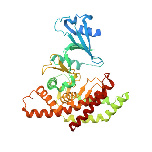
2CKO

Crystal structure of Human Choline Kinase alpha 2


Experimental Data Snapshot

  • Method: X-RAY DIFFRACTION
  • Resolution: 2.15 Å
  • R-Value Free: 0.256 
  • R-Value Work: 0.199 
  • R-Value Observed: 0.204 

Starting Model: experimental
View more details

wwPDB Validation   3D Report Full Report


This is version 1.3 of the entry. See complete history


Literature

Elucidation of Human Choline Kinase Crystal Structures in Complex with the Products Adp or Phosphocholine.

Malito, E.Sekulic, N.Too, W.C.Konrad, M.Lavie, A.

(2006) J Mol Biol 364: 136

  • DOI: https://doi.org/10.1016/j.jmb.2006.08.084
  • Primary Citation of Related Structures:  
    2CKO, 2CKP, 2CKQ

  • PubMed Abstract: 

    Choline kinase, responsible for the phosphorylation of choline to phosphocholine as the first step of the CDP-choline pathway for the biosynthesis of phosphatidylcholine, has been recognized as a new target for anticancer therapy. Crystal structures of human choline kinase in its apo, ADP and phosphocholine-bound complexes, respectively, reveal the molecular details of the substrate binding sites. ATP binds in a cavity where residues from both the N and C-terminal lobes contribute to form a cleft, while the choline-binding site constitutes a deep hydrophobic groove in the C-terminal domain with a rim composed of negatively charged residues. Upon binding of choline, the enzyme undergoes conformational changes independently affecting the N-terminal domain and the ATP-binding loop. From this structural analysis and comparison with other kinases, and from mutagenesis data on the homologous Caenorhabditis elegans choline kinase, a model of the ternary ADP.phosphocholine complex was built that reveals the molecular basis for the phosphoryl transfer activity of this enzyme.


  • Organizational Affiliation

    Department of Biochemistry and Molecular Genetics, University of Illinois at Chicago, Chicago, IL 60607, USA.


Macromolecules
Find similar proteins by:  (by identity cutoff)  |  3D Structure
Entity ID: 1
MoleculeChains Sequence LengthOrganismDetailsImage
CHOLINE KINASE ALPHA
A, B
390Homo sapiensMutation(s): 0 
EC: 2.7.1.32 (PDB Primary Data), 2.7.1.82 (UniProt)
UniProt & NIH Common Fund Data Resources
Find proteins for P35790 (Homo sapiens)
Explore P35790 
Go to UniProtKB:  P35790
PHAROS:  P35790
GTEx:  ENSG00000110721 
Entity Groups  
Sequence Clusters30% Identity50% Identity70% Identity90% Identity95% Identity100% Identity
UniProt GroupP35790
Sequence Annotations
Expand
  • Reference Sequence
Experimental Data & Validation

Experimental Data

  • Method: X-RAY DIFFRACTION
  • Resolution: 2.15 Å
  • R-Value Free: 0.256 
  • R-Value Work: 0.199 
  • R-Value Observed: 0.204 
  • Space Group: P 21 21 21
Unit Cell:
Length ( Å )Angle ( ˚ )
a = 56.18α = 90
b = 122.82β = 90
c = 132.17γ = 90
Software Package:
Software NamePurpose
REFMACrefinement
XDSdata reduction
PHASERphasing

Structure Validation

View Full Validation Report


Entry History 

Deposition Data

Revision History  (Full details and data files)

  • Version 1.0: 2006-10-04
    Type: Initial release
  • Version 1.1: 2011-05-08
    Changes: Version format compliance
  • Version 1.2: 2011-07-13
    Changes: Version format compliance
  • Version 1.3: 2023-12-13
    Changes: Data collection, Database references, Derived calculations, Other, Refinement description