2D0F

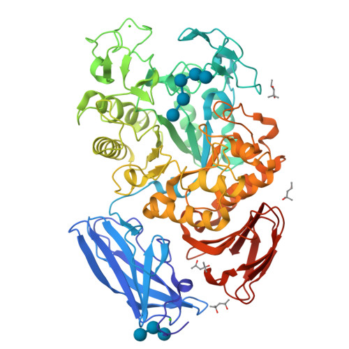
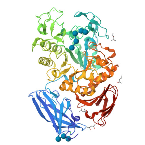
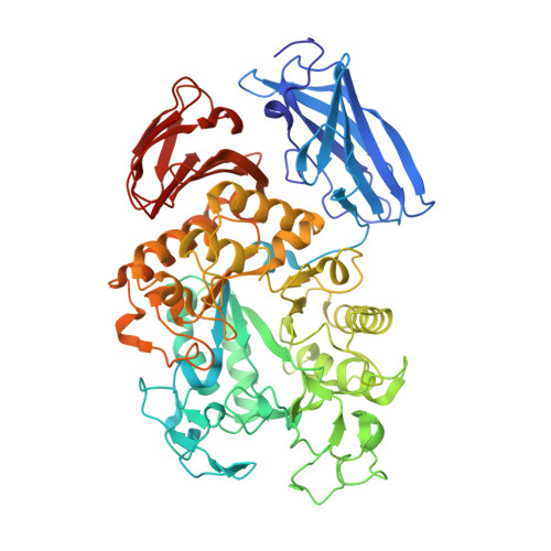
Crystal Structure of Thermoactinomyces vulgaris R-47 Alpha-Amylase 1 (TVAI) Mutant D356N complexed with P2, a pullulan model oligosaccharide


Experimental Data Snapshot

  • Method: X-RAY DIFFRACTION
  • Resolution: 2.08 Å
  • R-Value Free: 0.187 
  • R-Value Work: 0.154 
  • R-Value Observed: 0.157 

wwPDB Validation   3D Report Full Report


This is version 2.2 of the entry. See complete history


Literature

Complexes of Thermoactinomyces vulgaris R-47 alpha-amylase 1 and pullulan model oligossacharides provide new insight into the mechanism for recognizing substrates with alpha-(1,6) glycosidic linkages

Abe, A.Yoshida, H.Tonozuka, T.Sakano, Y.Kamitori, S.

(2005) FEBS J 272: 6145-6153

  • DOI: https://doi.org/10.1111/j.1742-4658.2005.05013.x
  • Primary Citation of Related Structures:  
    2D0F, 2D0G, 2D0H

  • PubMed Abstract: 

    Thermoactinomyces vulgaris R-47 alpha-amylase 1 (TVAI) has unique hydrolyzing activities for pullulan with sequence repeats of alpha-(1,4), alpha-(1,4), and alpha-(1,6) glycosidic linkages, as well as for starch. TVAI mainly hydrolyzes alpha-(1,4) glycosidic linkages to produce a panose, but it also hydrolyzes alpha-(1,6) glycosidic linkages with a lesser efficiency. X-ray structures of three complexes comprising an inactive mutant TVAI (D356N or D356N/E396Q) and a pullulan model oligosaccharide (P2; [Glc-alpha-(1,6)-Glc-alpha-(1,4)-Glc-alpha-(1,4)]2 or P5; [Glc-alpha-(1,6)-Glc-alpha-(1,4)-Glc-alpha-(1,4)]5) were determined. The complex D356N/P2 is a mimic of the enzyme/product complex in the main catalytic reaction of TVAI, and a structural comparison with Aspergillus oryzaealpha-amylase showed that the (-) subsites of TVAI are responsible for recognizing both starch and pullulan. D356N/E396Q/P2 and D356N/E396Q/P5 provided models of the enzyme/substrate complex recognizing the alpha-(1,6) glycosidic linkage at the hydrolyzing site. They showed that only subsites -1 and -2 at the nonreducing end of TVAI are effective in the hydrolysis of alpha-(1,6) glycosidic linkages, leading to weak interactions between substrates and the enzyme. Domain N of TVAI is a starch-binding domain acting as an anchor in the catalytic reaction of the enzyme. In this study, additional substrates were also found to bind to domain N, suggesting that domain N also functions as a pullulan-binding domain.


  • Organizational Affiliation

    Graduate School of Medicine, Kagawa University, Japan.


Macromolecules
Find similar proteins by:  (by identity cutoff)  |  3D Structure
Entity ID: 1
MoleculeChains Sequence LengthOrganismDetailsImage
alpha-amylase I637Thermoactinomyces vulgarisMutation(s): 1 
EC: 3.2.1.1 (PDB Primary Data), 3.2.1.135 (UniProt)
UniProt
Find proteins for Q60053 (Thermoactinomyces vulgaris)
Explore Q60053 
Go to UniProtKB:  Q60053
Entity Groups  
Sequence Clusters30% Identity50% Identity70% Identity90% Identity95% Identity100% Identity
UniProt GroupQ60053
Sequence Annotations
Expand
  • Reference Sequence
Oligosaccharides

Help

Entity ID: 2
MoleculeChains Length2D Diagram Glycosylation3D Interactions
alpha-D-glucopyranose-(1-4)-alpha-D-glucopyranose-(1-4)-alpha-D-glucopyranose-(1-6)-alpha-D-glucopyranose-(1-4)-beta-D-glucopyranose
B
5N/A
Glycosylation Resources
GlyTouCan:  G40634PP
GlyCosmos:  G40634PP
Entity ID: 3
MoleculeChains Length2D Diagram Glycosylation3D Interactions
alpha-D-glucopyranose-(1-4)-alpha-D-glucopyranose-(1-4)-beta-D-glucopyranose
C
3N/A
Glycosylation Resources
GlyTouCan:  G00395FS
GlyCosmos:  G00395FS
GlyGen:  G00395FS
Biologically Interesting Molecules (External Reference) 1 Unique
Experimental Data & Validation

Experimental Data

  • Method: X-RAY DIFFRACTION
  • Resolution: 2.08 Å
  • R-Value Free: 0.187 
  • R-Value Work: 0.154 
  • R-Value Observed: 0.157 
  • Space Group: C 1 2 1
Unit Cell:
Length ( Å )Angle ( ˚ )
a = 121.532α = 90
b = 50.429β = 104.3
c = 107.834γ = 90
Software Package:
Software NamePurpose
HKL-2000data collection
SCALEPACKdata scaling
CNSrefinement
HKL-2000data reduction
CNSphasing

Structure Validation

View Full Validation Report


Entry History 

Deposition Data

Revision History  (Full details and data files)

  • Version 1.0: 2006-07-11
    Type: Initial release
  • Version 1.1: 2008-04-30
    Changes: Version format compliance
  • Version 1.2: 2011-07-13
    Changes: Version format compliance
  • Version 2.0: 2020-07-29
    Type: Remediation
    Reason: Carbohydrate remediation
    Changes: Atomic model, Data collection, Derived calculations, Structure summary
  • Version 2.1: 2021-11-10
    Changes: Database references, Structure summary
  • Version 2.2: 2024-05-29
    Changes: Data collection