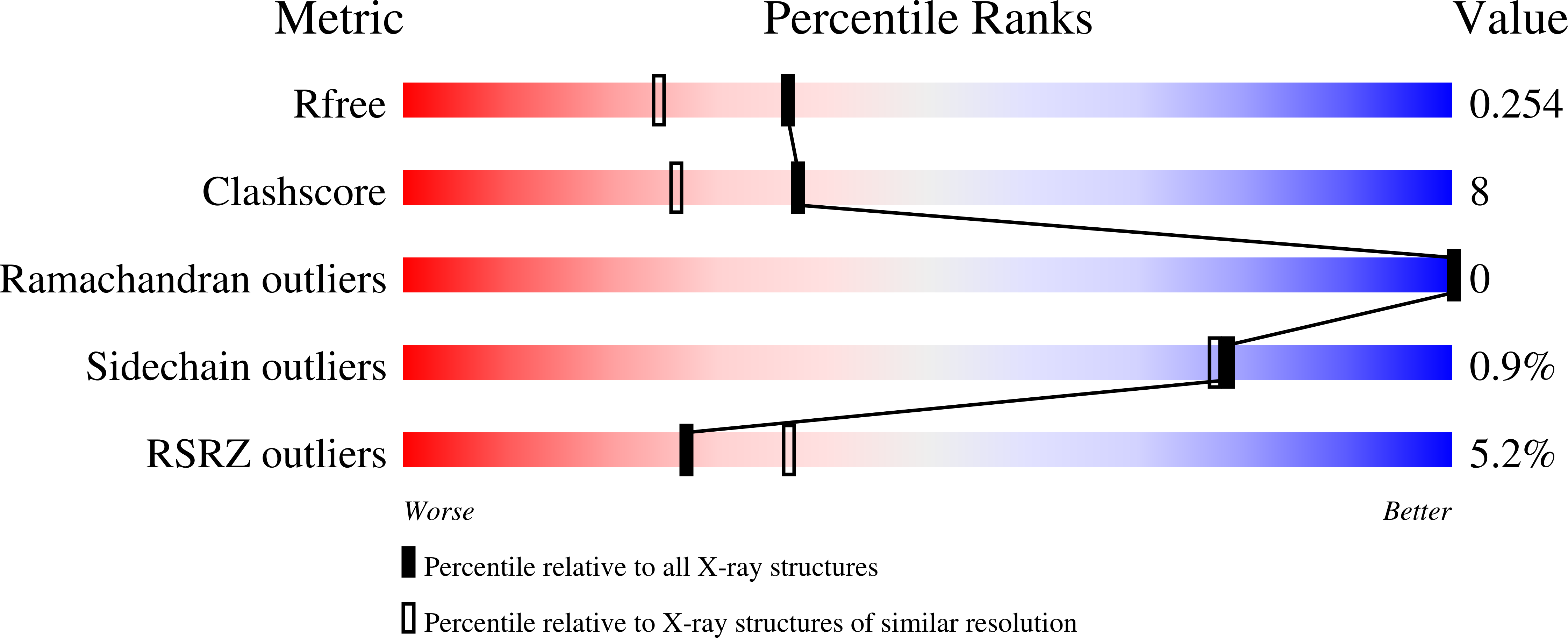
2DGM

Crystal structure of Escherichia coli GadB in complex with iodide


Experimental Data Snapshot

  • Method: X-RAY DIFFRACTION
  • Resolution: 1.95 Å
  • R-Value Free: 
    0.267 (Depositor), 0.250 (DCC) 
  • R-Value Work: 
    0.227 (Depositor), 0.220 (DCC) 

Starting Model: experimental
View more details

wwPDB Validation   3D Report Full Report


Ligand Structure Quality Assessment 

Created with Raphaël 2.3.0Worse 01 BetterLigand structure goodness of fit to experimental dataBest fitted PLPClick on this verticalbar to view details

This is version 1.3 of the entry. See complete history


Literature

Escherichia coli acid resistance: pH-sensing, activation by chloride and autoinhibition in GadB

Gut, H.Pennacchietti, E.John, R.A.Bossa, F.Capitani, G.De Biase, D.Gruetter, M.G.

(2006) EMBO J 25: 2643-2651

  • DOI: https://doi.org/10.1038/sj.emboj.7601107
  • Primary Citation of Related Structures:  
    2DGK, 2DGL, 2DGM

  • PubMed Abstract: 

    Escherichia coli and other enterobacteria exploit the H+ -consuming reaction catalysed by glutamate decarboxylase to survive the stomach acidity before reaching the intestine. Here we show that chloride, extremely abundant in gastric secretions, is an allosteric activator producing a 10-fold increase in the decarboxylase activity at pH 5.6. Cooperativity and sensitivity to chloride were lost when the N-terminal 14 residues, involved in the formation of two triple-helix bundles, were deleted by mutagenesis. X-ray structures, obtained in the presence of the substrate analogue acetate, identified halide-binding sites at the base of each N-terminal helix, showed how halide binding is responsible for bundle stability and demonstrated that the interconversion between active and inactive forms of the enzyme is a stepwise process. We also discovered an entirely novel structure of the cofactor pyridoxal 5'-phosphate (aldamine) to be responsible for the reversibly inactivated enzyme. Our results link the entry of chloride ions, via the H+/Cl- exchange activities of ClC-ec1, to the trigger of the acid stress response in the cell when the intracellular proton concentration has not yet reached fatal values.


  • Organizational Affiliation

    Biochemisches Institut der Universität Zürich, Zürich, Switzerland.


Macromolecules
Find similar proteins by:  (by identity cutoff)  |  3D Structure
Entity ID: 1
MoleculeChains Sequence LengthOrganismDetailsImage
Glutamate decarboxylase beta
A, B, C, D, E
466Escherichia coliMutation(s): 0 
Gene Names: gadB
EC: 4.1.1.15
UniProt
Find proteins for P69910 (Escherichia coli (strain K12))
Explore P69910 
Go to UniProtKB:  P69910
Entity Groups  
Sequence Clusters30% Identity50% Identity70% Identity90% Identity95% Identity100% Identity
UniProt GroupP69910
Sequence Annotations
Expand
  • Reference Sequence
Small Molecules
Ligands 5 Unique
IDChains Name / Formula / InChI Key2D Diagram3D Interactions
PLP
Query on PLP

Download Ideal Coordinates CCD File 
CA [auth C]
CB [auth F]
KA [auth D]
M [auth A]
TA [auth E]
PYRIDOXAL-5'-PHOSPHATE
C8 H10 N O6 P
NGVDGCNFYWLIFO-UHFFFAOYSA-N
IOD
Query on IOD

Download Ideal Coordinates CCD File 
AA [auth C]
AB [auth F]
BA [auth C]
BB [auth F]
EA [auth D]
IODIDE ION
I
XMBWDFGMSWQBCA-UHFFFAOYSA-M
PEG
Query on PEG

Download Ideal Coordinates CCD File 
WA [auth E]DI(HYDROXYETHYL)ETHER
C4 H10 O3
MTHSVFCYNBDYFN-UHFFFAOYSA-N
ACY
Query on ACY

Download Ideal Coordinates CCD File 
VA [auth E]ACETIC ACID
C2 H4 O2
QTBSBXVTEAMEQO-UHFFFAOYSA-N
FMT
Query on FMT

Download Ideal Coordinates CCD File 
DA [auth C]
DB [auth F]
EB [auth F]
FB [auth F]
GB [auth F]
FORMIC ACID
C H2 O2
BDAGIHXWWSANSR-UHFFFAOYSA-N
Experimental Data & Validation

Experimental Data

  • Method: X-RAY DIFFRACTION
  • Resolution: 1.95 Å
  • R-Value Free:  0.267 (Depositor), 0.250 (DCC) 
  • R-Value Work:  0.227 (Depositor), 0.220 (DCC) 
Space Group: P 1
Unit Cell:
Length ( Å )Angle ( ˚ )
a = 91.336α = 76.65
b = 91.804β = 76.94
c = 93.914γ = 78.04
Software Package:
Software NamePurpose
DENZOdata reduction
SCALEPACKdata scaling
AMoREphasing
CNSrefinement

Structure Validation

View Full Validation Report


Ligand Structure Quality Assessment 

Created with Raphaël 2.3.0Worse 01 BetterLigand structure goodness of fit to experimental dataBest fitted PLPClick on this verticalbar to view details

Entry History 

Deposition Data

Revision History  (Full details and data files)

  • Version 1.0: 2006-06-20
    Type: Initial release
  • Version 1.1: 2008-04-30
    Changes: Version format compliance
  • Version 1.2: 2011-07-13
    Changes: Non-polymer description, Version format compliance
  • Version 1.3: 2023-10-25
    Changes: Data collection, Database references, Derived calculations, Refinement description