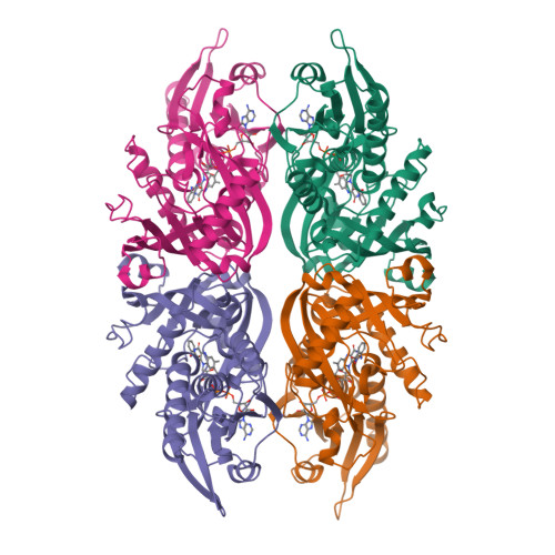
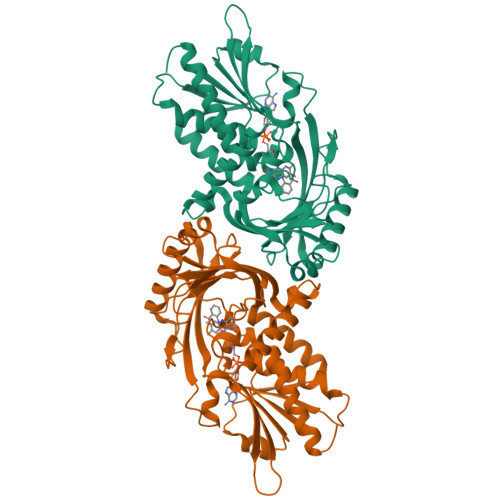
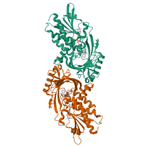
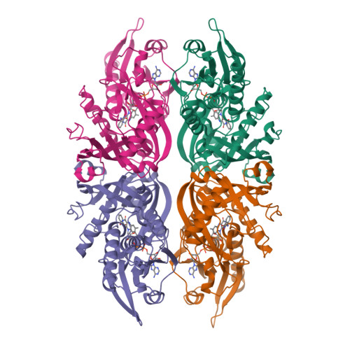
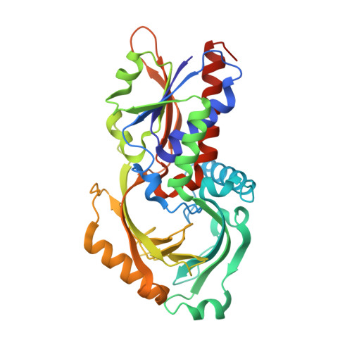
2E4A

Crystal Structure of Human D-Amino Acid Oxidase in complex with o-aminobenzoate


Experimental Data Snapshot

  • Method: X-RAY DIFFRACTION
  • Resolution: 2.60 Å
  • R-Value Free: 0.244 
  • R-Value Work: 0.210 
  • R-Value Observed: 0.212 

Starting Model: experimental
View more details

wwPDB Validation   3D Report Full Report


Ligand Structure Quality Assessment 


This is version 2.0 of the entry. See complete history


Literature

Structural basis of d-DOPA oxidation by d-amino acid oxidase: Alternative pathway for dopamine biosynthesis.

Kawazoe, T.Tsuge, H.Imagawa, T.Aki, K.Kuramitsu, S.Fukui, K.

(2007) Biochem Biophys Res Commun 355: 385-391

  • DOI: https://doi.org/10.1016/j.bbrc.2007.01.181
  • Primary Citation of Related Structures:  
    2E48, 2E49, 2E4A, 2E82

  • PubMed Abstract: 

    D-amino acid oxidase (DAO) degrades the gliotransmitter D-serine, a potent endogenous ligand of N-methyl-D-aspartate type glutamate receptors. It also has been suggested that D-DOPA, the stereoisomer of L-DOPA, is oxidized by DAO and then converted to dopamine via an alternative biosynthetic pathway. Here, we provide direct crystallographic evidence that D-DOPA is readily fitted into the active site of human DAO, where it is oxidized by the enzyme. Moreover, our kinetic data show that the maximal velocity for oxidation of D-DOPA is much greater than for D-serine, which strongly supports the proposed alternative pathway for dopamine biosynthesis in the treatment of Parkinson's disease. In addition, determination of the structures of human DAO in various states revealed that the conformation of the hydrophobic VAAGL stretch (residues 47-51) to be uniquely stable in the human enzyme, which provides a structural basis for the unique kinetic features of human DAO.


  • Organizational Affiliation

    The Institute for Enzyme Research, The University of Tokushima, 3-18-15 Kuramoto, Tokushima 770-8503, Japan.


Macromolecules
Find similar proteins by:  (by identity cutoff)  |  3D Structure
Entity ID: 1
MoleculeChains Sequence LengthOrganismDetailsImage
D-amino-acid oxidase
A, B, C, D
347Homo sapiensMutation(s): 0 
EC: 1.4.3.3
UniProt & NIH Common Fund Data Resources
Find proteins for P14920 (Homo sapiens)
Explore P14920 
Go to UniProtKB:  P14920
PHAROS:  P14920
GTEx:  ENSG00000110887 
Entity Groups  
Sequence Clusters30% Identity50% Identity70% Identity90% Identity95% Identity100% Identity
UniProt GroupP14920
Sequence Annotations
Expand
  • Reference Sequence
Small Molecules
Ligands 2 Unique
IDChains Name / Formula / InChI Key2D Diagram3D Interactions
FAD
Query on FAD

Download Ideal Coordinates CCD File 
E [auth A],
G [auth B],
I [auth C],
K [auth D]
FLAVIN-ADENINE DINUCLEOTIDE
C27 H33 N9 O15 P2
VWWQXMAJTJZDQX-UYBVJOGSSA-N
BE2
Query on BE2

Download Ideal Coordinates CCD File 
F [auth A],
H [auth B],
J [auth C],
L [auth D]
2-AMINOBENZOIC ACID
C7 H7 N O2
RWZYAGGXGHYGMB-UHFFFAOYSA-N
Experimental Data & Validation

Experimental Data

  • Method: X-RAY DIFFRACTION
  • Resolution: 2.60 Å
  • R-Value Free: 0.244 
  • R-Value Work: 0.210 
  • R-Value Observed: 0.212 
  • Space Group: P 21 21 2
Unit Cell:
Length ( Å )Angle ( ˚ )
a = 151.392α = 90
b = 185.042β = 90
c = 51.577γ = 90
Software Package:
Software NamePurpose
REFMACrefinement
HKL-2000data scaling
MOLREPphasing

Structure Validation

View Full Validation Report


Ligand Structure Quality Assessment 


Entry History 

Deposition Data

Revision History  (Full details and data files)

  • Version 1.0: 2007-03-06
    Type: Initial release
  • Version 1.1: 2008-04-30
    Changes: Version format compliance
  • Version 1.2: 2011-07-13
    Changes: Version format compliance
  • Version 1.3: 2023-10-25
    Changes: Data collection, Database references, Derived calculations, Refinement description
  • Version 2.0: 2023-11-15
    Changes: Atomic model, Data collection