2I56 | pdb_00002i56

Crystal structure of L-Rhamnose Isomerase from Pseudomonas stutzeri with L-Rhamnose


Experimental Data Snapshot

  • Method: X-RAY DIFFRACTION
  • Resolution: 1.97 Å
  • R-Value Free: 
    0.205 (Depositor), 0.200 (DCC) 
  • R-Value Work: 
    0.170 (Depositor), 0.170 (DCC) 
  • R-Value Observed: 
    0.170 (Depositor) 

Starting Model: experimental
View more details

wwPDB Validation   3D Report Full Report


Ligand Structure Quality Assessment 

Created with Raphaël 2.3.0Worse 01 BetterLigand structure goodness of fit to experimental dataBest fitted RNSClick on this verticalbar to view details

This is version 1.4 of the entry. See complete history


Literature

The Structures of l-Rhamnose Isomerase from Pseudomonas stutzeri in Complexes with l-Rhamnose and d-Allose Provide Insights into Broad Substrate Specificity

Yoshida, H.Yamada, M.Ohyama, Y.Takada, G.Izumori, K.Kamitori, S.

(2007) J Mol Biology 365: 1505-1516

  • DOI: https://doi.org/10.1016/j.jmb.2006.11.004
  • Primary Citation of Related Structures:  
    2HCV, 2I56, 2I57

  • PubMed Abstract: 

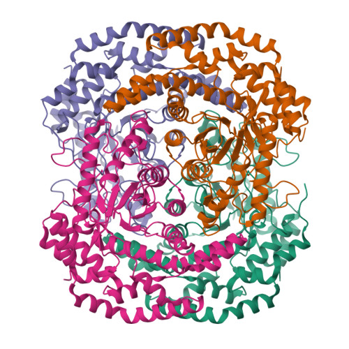
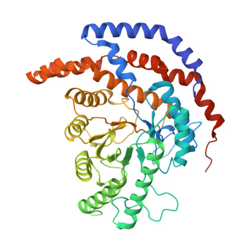
    Pseudomonas stutzeri L-rhamnose isomerase (P. stutzeri L-RhI) can efficiently catalyze the isomerization between various aldoses and ketoses, showing a broad substrate specificity compared to L-RhI from Escherichia coli (E. coli L-RhI). To understand the relationship between structure and substrate specificity, the crystal structures of P. stutzeri L-RhI alone and in complexes with L-rhamnose and D-allose which has different configurations of C4 and C5 from L-rhamnose, were determined at a resolution of 2.0 A, 1.97 A, and 1.97 A, respectively. P. stutzeri L-RhI has a large domain with a (beta/alpha)(8) barrel fold and an additional small domain composed of seven alpha-helices, forming a homo tetramer, as found in E. coli L-RhI and D-xylose isomerases (D-XIs) from various microorganisms. The beta1-alpha1 loop (Gly60-Arg76) of P. stutzeri L-RhI is involved in the substrate binding of a neighbouring molecule, as found in D-XIs, while in E. coli L-RhI, the corresponding beta1-alpha1 loop is extended (Asp52-Arg78) and covers the substrate-binding site of the same molecule. The complex structures of P. stutzeri L-RhI with L-rhamnose and D-allose show that both substrates are nicely fitted to the substrate-binding site. The part of the substrate-binding site interacting with the substrate at the 1, 2, and 3 positions is equivalent to E. coli L-RhI, and the other part interacting with the 4, 5, and 6 positions is similar to D-XI. In E. coli L-RhI, the beta1-alpha1 loop creates an unique hydrophobic pocket at the the 4, 5, and 6 positions, leading to the strictly recognition of L-rhamnose as the most suitable substrate, while in P. stutzeri L-RhI, there is no corresponding hydrophobic pocket where Phe66 from a neighbouring molecule merely forms hydrophobic interactions with the substrate, leading to the loose substrate recognition at the 4, 5, and 6 positions.


  • Organizational Affiliation

    Molecular Structure Research Group, Information Technology Center and Faculty of Medicine, Kagawa University, 1750-1 Ikenobe, Miki-cho, Kita-gun, Kagawa 761-0793, Japan.


Macromolecules
Find similar proteins by:  (by identity cutoff)  |  3D Structure
Entity ID: 1
MoleculeChains Sequence LengthOrganismDetailsImage
L-rhamnose isomerase
A, B, C, D
438Stutzerimonas stutzeriMutation(s): 1 
EC: 5.3.1.14
UniProt
Find proteins for Q75WH8 (Stutzerimonas stutzeri)
Explore Q75WH8 
Go to UniProtKB:  Q75WH8
Entity Groups  
Sequence Clusters30% Identity50% Identity70% Identity90% Identity95% Identity100% Identity
UniProt GroupQ75WH8
Sequence Annotations
Expand
  • Reference Sequence
Small Molecules
Ligands 2 Unique
IDChains Name / Formula / InChI Key2D Diagram3D Interactions
RNS
Query on RNS

Download Ideal Coordinates CCD File 
E [auth A],
H [auth B],
K [auth C],
N [auth D]
L-RHAMNOSE
C6 H12 O5
PNNNRSAQSRJVSB-BXKVDMCESA-N
ZN
Query on ZN

Download Ideal Coordinates CCD File 
F [auth A]
G [auth A]
I [auth B]
J [auth B]
L [auth C]
ZINC ION
Zn
PTFCDOFLOPIGGS-UHFFFAOYSA-N
Experimental Data & Validation

Experimental Data

  • Method: X-RAY DIFFRACTION
  • Resolution: 1.97 Å
  • R-Value Free:  0.205 (Depositor), 0.200 (DCC) 
  • R-Value Work:  0.170 (Depositor), 0.170 (DCC) 
  • R-Value Observed: 0.170 (Depositor) 
Space Group: P 1 21 1
Unit Cell:
Length ( Å )Angle ( ˚ )
a = 74.616α = 90
b = 104.757β = 107.96
c = 114.292γ = 90
Software Package:
Software NamePurpose
CNSrefinement
ADSCdata collection
HKL-2000data reduction
HKL-2000data scaling
MOLREPphasing

Structure Validation

View Full Validation Report


Ligand Structure Quality Assessment 

Created with Raphaël 2.3.0Worse 01 BetterLigand structure goodness of fit to experimental dataBest fitted RNSClick on this verticalbar to view details

Entry History 

Deposition Data

Revision History  (Full details and data files)

  • Version 1.0: 2006-12-19
    Type: Initial release
  • Version 1.1: 2008-05-01
    Changes: Version format compliance
  • Version 1.2: 2011-07-13
    Changes: Version format compliance
  • Version 1.3: 2021-11-10
    Changes: Database references, Derived calculations
  • Version 1.4: 2024-04-03
    Changes: Data collection, Refinement description