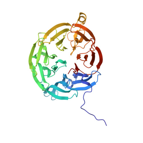
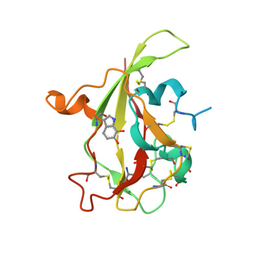
2IUQ

CRYSTAL STRUCTURE OF DITHIONITE-REDUCED AROMATIC AMINE DEHYDROGENASE (AADH) FROM ALCALIGENES FAECALIS IN COMPLEX WITH TRYPTAMINE


Experimental Data Snapshot

  • Method: X-RAY DIFFRACTION
  • Resolution: 1.50 Å
  • R-Value Free: 0.190 
  • R-Value Work: 0.157 
  • R-Value Observed: 0.158 

wwPDB Validation   3D Report Full Report


Ligand Structure Quality Assessment 


This is version 1.4 of the entry. See complete history


Literature

Atomic Level Insight Into the Oxidative Half-Reaction of Aromatic Amine Dehydrogenase.

Roujeinikova, A.Scrutton, N.Leys, D.

(2006) J Biol Chem 281: 40264

  • DOI: https://doi.org/10.1074/jbc.M605559200
  • Primary Citation of Related Structures:  
    2HXC, 2IUP, 2IUQ, 2IUR, 2IUV

  • PubMed Abstract: 

    The quinoprotein aromatic amine dehydrogenase (AADH) uses a covalently bound tryptophan tryptophylquinone (TTQ) cofactor to oxidatively deaminate primary aromatic amines. Recent crystal structures have provided insight into the reductive half-reaction. In contrast, no atomic details are available for the oxidative half-reaction. The TTQ O7 hydroxyl group is protonated during reduction, but it is unclear how this proton can be removed during the oxidative half-reaction. Furthermore, compared with the electron transfer from the N-quinol form, electron transfer from the non-physiological O-quinol form to azurin is significantly slower. Here we report crystal structures of the O-quinol, N-quinol, and N-semiquinone forms of AADH. A comparison of oxidized and substrate reduced AADH species reveals changes in the TTQ-containing subunit, extending from residues in the immediate vicinity of the N-quinol to the putative azurin docking site, suggesting a mechanism whereby TTQ redox state influences interprotein electron transfer. In contrast, chemical reduction of the TTQ center has no significant effect on protein conformation. Furthermore, structural reorganization upon substrate reduction places a water molecule near TTQ O7 where it can act as proton acceptor. The structure of the N-semiquinone, however, is essentially similar to oxidized AADH. Surprisingly, in the presence of substrate a covalent N-semiquinone substrate adduct is observed. To our knowledge this is the first detailed insight into a complex, branching mechanism of quinone oxidation where significant structural reorganization upon reduction of the quinone center directly influences formation of the electron transfer complex and nature of the electron transfer process.


  • Organizational Affiliation

    Manchester Interdisciplinary Biocentre, University of Manchester, Manchester M1 7DN, United Kingdom.


Macromolecules
Find similar proteins by:  (by identity cutoff)  |  3D Structure
Entity ID: 1
MoleculeChains Sequence LengthOrganismDetailsImage
AROMATIC AMINE DEHYDROGENASE ALPHA SUBUNIT
A, B
361Alcaligenes faecalisMutation(s): 0 
EC: 1.4.99.4 (PDB Primary Data), 1.4.9.2 (UniProt)
UniProt
Find proteins for P84888 (Alcaligenes faecalis)
Explore P84888 
Go to UniProtKB:  P84888
Entity Groups  
Sequence Clusters30% Identity50% Identity70% Identity90% Identity95% Identity100% Identity
UniProt GroupP84888
Sequence Annotations
Expand
  • Reference Sequence
Find similar proteins by:  (by identity cutoff)  |  3D Structure
Entity ID: 2
MoleculeChains Sequence LengthOrganismDetailsImage
AROMATIC AMINE DEHYDROGENASE BETA SUBUNITC [auth D],
D [auth H]
135Alcaligenes faecalisMutation(s): 0 
EC: 1.4.99.4 (PDB Primary Data), 1.4.9.2 (UniProt)
UniProt
Find proteins for P84887 (Alcaligenes faecalis)
Explore P84887 
Go to UniProtKB:  P84887
Entity Groups  
Sequence Clusters30% Identity50% Identity70% Identity90% Identity95% Identity100% Identity
UniProt GroupP84887
Sequence Annotations
Expand
  • Reference Sequence
Small Molecules
Modified Residues  1 Unique
IDChains TypeFormula2D DiagramParent
TRQ
Query on TRQ
C [auth D],
D [auth H]
L-PEPTIDE LINKINGC11 H10 N2 O4TRP
Experimental Data & Validation

Experimental Data

  • Method: X-RAY DIFFRACTION
  • Resolution: 1.50 Å
  • R-Value Free: 0.190 
  • R-Value Work: 0.157 
  • R-Value Observed: 0.158 
  • Space Group: P 1 21 1
Unit Cell:
Length ( Å )Angle ( ˚ )
a = 71.139α = 90
b = 89.028β = 90.1
c = 80.727γ = 90
Software Package:
Software NamePurpose
REFMACrefinement
DENZOdata reduction
SCALEPACKdata scaling

Structure Validation

View Full Validation Report


Ligand Structure Quality Assessment 


Entry History 

Deposition Data

Revision History  (Full details and data files)

  • Version 1.0: 2006-09-20
    Type: Initial release
  • Version 1.1: 2011-05-08
    Changes: Version format compliance
  • Version 1.2: 2011-07-13
    Changes: Version format compliance
  • Version 1.3: 2019-01-30
    Changes: Data collection, Experimental preparation
  • Version 1.4: 2019-02-06
    Changes: Data collection, Experimental preparation