2PHN

Crystal structure of an amide bond forming F420-gamma glutamyl ligase from Archaeoglobus fulgidus


Experimental Data Snapshot

  • Method: X-RAY DIFFRACTION
  • Resolution: 1.35 Å
  • R-Value Free: 0.190 
  • R-Value Work: 0.160 
  • R-Value Observed: 0.162 

Starting Model: experimental
View more details

wwPDB Validation   3D Report Full Report


Ligand Structure Quality Assessment 


This is version 1.5 of the entry. See complete history


Literature

Structure of an Amide Bond Forming F(420):gammagamma-glutamyl Ligase from Archaeoglobus Fulgidus - A Member of a New Family of Non-ribosomal Peptide Synthases.

Nocek, B.Evdokimova, E.Proudfoot, M.Kudritska, M.Grochowski, L.L.White, R.H.Savchenko, A.Yakunin, A.F.Edwards, A.Joachimiak, A.

(2007) J Mol Biol 372: 456-469

  • DOI: https://doi.org/10.1016/j.jmb.2007.06.063
  • Primary Citation of Related Structures:  
    2G9I, 2PHN

  • PubMed Abstract: 

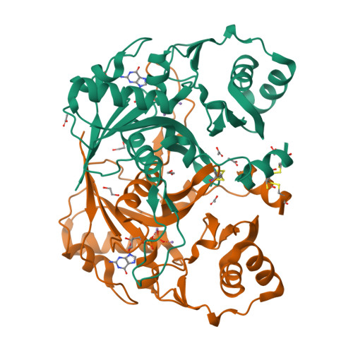
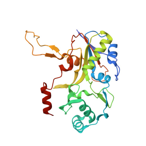
    F(420) is a flavin-like redox-active coenzyme commonly used by archaea and some eubacteria in a variety of biochemical reactions in methanogenesis, the formation of secondary metabolites, the degradation of nitroaromatic compounds, activation of nitroimidazofurans, and F(420)-dependent photolysis in DNA repair. Coenzyme F(420)-2 biosynthesis from 7,8-didemethyl-8-hydroxy-5-deazariboflavin (Fo) and lactaldehyde involves six enzymatic steps and five proteins (CofA, CofB, CofC, CofD, and CofE). CofE, a F(420)-0:gamma-glutamyl ligase, is responsible for the last two enzymatic steps; it catalyses the GTP-dependent addition of two L-glutamate residues to F(420)-0 to form F(420)-2. CofE is found in archaea, the aerobic actinomycetes, and cyanobacteria. Here, we report the first crystal structure of the apo-F(420)-0:gamma-glutamyl ligase (CofE-AF) from Archaeoglobus fulgidus and its complex with GDP at 2.5 A and 1.35 A resolution, respectively. The structure of CofE-AF reveals a novel protein fold with an intertwined, butterfly-like dimer formed by two-domain monomers. GDP and Mn(2+) are bound within the putative active site in a large groove at the dimer interface. We show that the enzyme adds a glutamate residue to both F(420)-0 and F(420)-1 in two distinct steps. CofE represents the first member of a new structural family of non-ribosomal peptide synthases.


  • Organizational Affiliation

    Midwest Center for Structural Genomics and Structural Biology Center, Biosciences, Argonne National Laboratory, Argonne, IL 60439, USA.


Macromolecules
Find similar proteins by:  (by identity cutoff)  |  3D Structure
Entity ID: 1
MoleculeChains Sequence LengthOrganismDetailsImage
F420-0:gamma-glutamyl ligase
A, B
254Archaeoglobus fulgidus DSM 4304Mutation(s): 0 
Gene Names: cofEAF_2256
EC: 6.3.2
UniProt
Find proteins for O28028 (Archaeoglobus fulgidus (strain ATCC 49558 / DSM 4304 / JCM 9628 / NBRC 100126 / VC-16))
Explore O28028 
Go to UniProtKB:  O28028
Entity Groups  
Sequence Clusters30% Identity50% Identity70% Identity90% Identity95% Identity100% Identity
UniProt GroupO28028
Sequence Annotations
Expand
  • Reference Sequence
Small Molecules
Ligands 5 Unique
IDChains Name / Formula / InChI Key2D Diagram3D Interactions
GDP
Query on GDP

Download Ideal Coordinates CCD File 
H [auth A],
Q [auth B]
GUANOSINE-5'-DIPHOSPHATE
C10 H15 N5 O11 P2
QGWNDRXFNXRZMB-UUOKFMHZSA-N
GOL
Query on GOL

Download Ideal Coordinates CCD File 
M [auth A]GLYCEROL
C3 H8 O3
PEDCQBHIVMGVHV-UHFFFAOYSA-N
EDO
Query on EDO

Download Ideal Coordinates CCD File 
I [auth A],
J [auth A],
K [auth A],
L [auth A]
1,2-ETHANEDIOL
C2 H6 O2
LYCAIKOWRPUZTN-UHFFFAOYSA-N
ACT
Query on ACT

Download Ideal Coordinates CCD File 
E [auth A],
F [auth A],
G [auth A],
P [auth B]
ACETATE ION
C2 H3 O2
QTBSBXVTEAMEQO-UHFFFAOYSA-M
MN
Query on MN

Download Ideal Coordinates CCD File 
C [auth A],
D [auth A],
N [auth B],
O [auth B]
MANGANESE (II) ION
Mn
WAEMQWOKJMHJLA-UHFFFAOYSA-N
Experimental Data & Validation

Experimental Data

  • Method: X-RAY DIFFRACTION
  • Resolution: 1.35 Å
  • R-Value Free: 0.190 
  • R-Value Work: 0.160 
  • R-Value Observed: 0.162 
  • Space Group: P 41 21 2
Unit Cell:
Length ( Å )Angle ( ˚ )
a = 98.367α = 90
b = 98.367β = 90
c = 94.347γ = 90
Software Package:
Software NamePurpose
REFMACrefinement
SBC-Collectdata collection
HKL-3000data reduction
HKL-3000data scaling
MOLREPphasing

Structure Validation

View Full Validation Report


Ligand Structure Quality Assessment 


Entry History 

Revision History  (Full details and data files)

  • Version 1.0: 2007-05-15
    Type: Initial release
  • Version 1.1: 2008-05-01
    Changes: Version format compliance
  • Version 1.2: 2011-07-13
    Changes: Non-polymer description, Version format compliance
  • Version 1.3: 2011-10-19
    Changes: Derived calculations
  • Version 1.4: 2023-08-30
    Changes: Data collection, Database references, Derived calculations, Refinement description
  • Version 1.5: 2023-12-27
    Changes: Derived calculations