2PNU

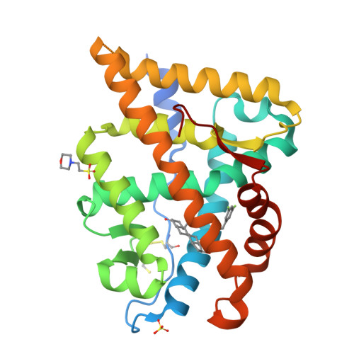
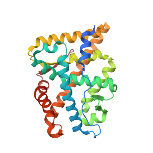
Crystal structure of human androgen receptor ligand-binding domain in complex with EM-5744


Experimental Data Snapshot

  • Method: X-RAY DIFFRACTION
  • Resolution: 1.65 Å
  • R-Value Free: 0.205 
  • R-Value Work: 0.183 
  • R-Value Observed: 0.183 

Starting Model: experimental
View more details

wwPDB Validation   3D Report Full Report


Ligand Structure Quality Assessment 


This is version 1.3 of the entry. See complete history


Literature

Structural Characterization of the Human Androgen Receptor Ligand-binding Domain Complexed with EM5744, a Rationally Designed Steroidal Ligand Bearing a Bulky Chain Directed toward Helix 12.

Cantin, L.Faucher, F.Couture, J.F.de Jesus-Tran, K.P.Legrand, P.Ciobanu, L.C.Frechette, Y.Labrecque, R.Singh, S.M.Labrie, F.Breton, R.

(2007) J Biol Chem 282: 30910-30919

  • DOI: https://doi.org/10.1074/jbc.M705524200
  • Primary Citation of Related Structures:  
    2PNU

  • PubMed Abstract: 

    Antiandrogens are commonly used to treat androgen-dependent disorders. The currently used drugs unfortunately possess very weak affinity for the human AR (hAR), thus indicating the need to develop new high-affinity steroidal antiandrogens. Our compounds are specially designed to impede repositioning of the mobile carboxyl-terminal helix 12, which blocks the ligand-dependent transactivation function (AF-2) located in the AR ligand-binding domain (ARLBD). Using crystal structures of the hARLBD, we first found that H12 could be directly reached from the ligand-binding pocket (LBP) by a chain positioned on the C18 atom of an androgen steroid nucleus. A set of 5alpha-dihydrotestosterone-derived molecules bearing various C18 chains were thus synthesized and tested for their capacity to bind hAR and act as antagonists. Although most of those having very high affinity for hAR were agonists, several very potent antagonists were obtained, confirming the structural importance of the C18 chain. To understand the role of the C18 chain in their agonistic/antagonistic properties, the structure of the hARLBD complexed with one of these agonists, EM5744, was determined at a 1.65-A resolution. We have identified new interactions involving Gln(738), Met(742), and His(874) that explain both the high affinity of this compound and the inability of its bulky chain to prevent the repositioning of H12. This structural information will be helpful to refine the structure of the chains placed on the C18 atom to obtain efficient H12-directed steroidal antiandrogens.


  • Organizational Affiliation

    Oncology and Molecular Endocrinology Research Center, Laval University Medical Center (CHUL) and Laval University, Québec G1V 4G2, Canada.


Macromolecules
Find similar proteins by:  (by identity cutoff)  |  3D Structure
Entity ID: 1
MoleculeChains Sequence LengthOrganismDetailsImage
Androgen receptor266Homo sapiensMutation(s): 0 
Gene Names: ARDHTRNR3C4
UniProt & NIH Common Fund Data Resources
Find proteins for P10275 (Homo sapiens)
Explore P10275 
Go to UniProtKB:  P10275
PHAROS:  P10275
GTEx:  ENSG00000169083 
Entity Groups  
Sequence Clusters30% Identity50% Identity70% Identity90% Identity95% Identity100% Identity
UniProt GroupP10275
Sequence Annotations
Expand
  • Reference Sequence
Small Molecules
Ligands 4 Unique
IDChains Name / Formula / InChI Key2D Diagram3D Interactions
ENM
Query on ENM

Download Ideal Coordinates CCD File 
C [auth A](5S,8R,9S,10S,13R,14S,17S)-13-{2-[(3,5-DIFLUOROBENZYL)OXY]ETHYL}-17-HYDROXY-10-METHYLHEXADECAHYDRO-3H-CYCLOPENTA[A]PHENANTHREN-3-ONE
C27 H36 F2 O3
AJODXHGZHBERGJ-JLYQOUBASA-N
MES
Query on MES

Download Ideal Coordinates CCD File 
E [auth A]2-(N-MORPHOLINO)-ETHANESULFONIC ACID
C6 H13 N O4 S
SXGZJKUKBWWHRA-UHFFFAOYSA-N
DTT
Query on DTT

Download Ideal Coordinates CCD File 
D [auth A]2,3-DIHYDROXY-1,4-DITHIOBUTANE
C4 H10 O2 S2
VHJLVAABSRFDPM-IMJSIDKUSA-N
SO4
Query on SO4

Download Ideal Coordinates CCD File 
B [auth A]SULFATE ION
O4 S
QAOWNCQODCNURD-UHFFFAOYSA-L
Experimental Data & Validation

Experimental Data

  • Method: X-RAY DIFFRACTION
  • Resolution: 1.65 Å
  • R-Value Free: 0.205 
  • R-Value Work: 0.183 
  • R-Value Observed: 0.183 
  • Space Group: P 21 21 21
Unit Cell:
Length ( Å )Angle ( ˚ )
a = 53.65α = 90
b = 66.36β = 90
c = 70.78γ = 90
Software Package:
Software NamePurpose
XSCALEdata processing
CNSrefinement
PDB_EXTRACTdata extraction
ADSCdata collection
XDSdata reduction
XSCALEdata scaling

Structure Validation

View Full Validation Report


Ligand Structure Quality Assessment 


Entry History 

Deposition Data

Revision History  (Full details and data files)

  • Version 1.0: 2007-09-11
    Type: Initial release
  • Version 1.1: 2011-07-13
    Changes: Version format compliance
  • Version 1.2: 2023-08-30
    Changes: Data collection, Database references, Derived calculations, Refinement description
  • Version 1.3: 2024-10-16
    Changes: Structure summary