2Q5O

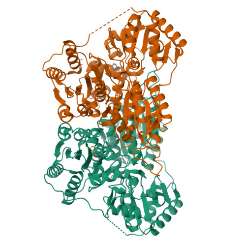
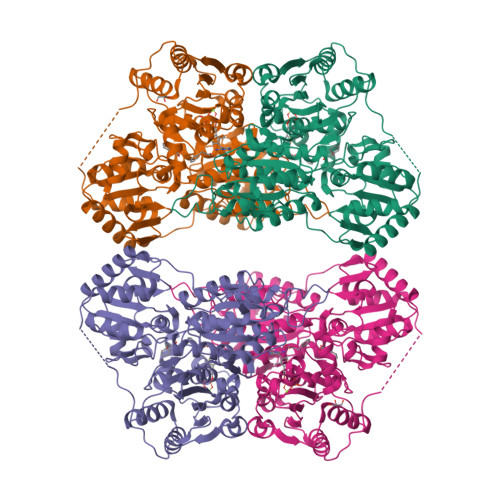
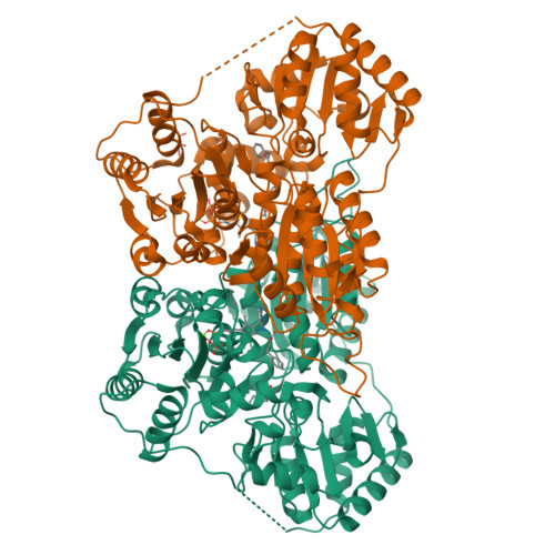
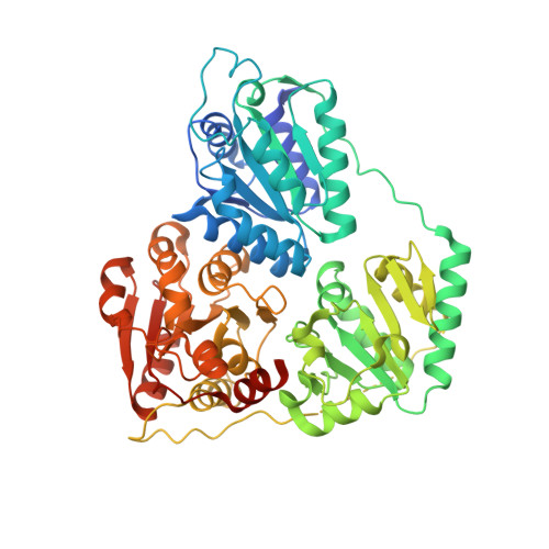
X-ray structure of phenylpyruvate decarboxylase in complex with 3-deaza-ThDP and phenylpyruvate


Experimental Data Snapshot

  • Method: X-RAY DIFFRACTION
  • Resolution: 2.15 Å
  • R-Value Free: 0.240 
  • R-Value Work: 0.182 
  • R-Value Observed: 0.182 

Starting Model: experimental
View more details

wwPDB Validation   3D Report Full Report


Ligand Structure Quality Assessment 


This is version 1.2 of the entry. See complete history


Literature

Molecular mechanism of allosteric substrate activation in a thiamine diphosphate-dependent decarboxylase.

Versees, W.Spaepen, S.Wood, M.D.Leeper, F.J.Vanderleyden, J.Steyaert, J.

(2007) J Biol Chem 282: 35269-35278

  • DOI: https://doi.org/10.1074/jbc.M706048200
  • Primary Citation of Related Structures:  
    2Q5J, 2Q5L, 2Q5O, 2Q5Q

  • PubMed Abstract: 

    Thiamine diphosphate-dependent enzymes are involved in a wide variety of metabolic pathways. The molecular mechanism behind active site communication and substrate activation, observed in some of these enzymes, has since long been an area of debate. Here, we report the crystal structures of a phenylpyruvate decarboxylase in complex with its substrates and a covalent reaction intermediate analogue. These structures reveal the regulatory site and unveil the mechanism of allosteric substrate activation. This signal transduction relies on quaternary structure reorganizations, domain rotations, and a pathway of local conformational changes that are relayed from the regulatory site to the active site. The current findings thus uncover the molecular mechanism by which the binding of a substrate in the regulatory site is linked to the mounting of the catalytic machinery in the active site in this thiamine diphosphate-dependent enzyme.


  • Organizational Affiliation

    Department of Ultrastructure, Vrije Universiteit Brussel, Pleinlaan 2, B-1050 Brussels, Belgium. wversees@vub.ac.be


Macromolecules
Find similar proteins by:  (by identity cutoff)  |  3D Structure
Entity ID: 1
MoleculeChains Sequence LengthOrganismDetailsImage
Phenylpyruvate decarboxylase
A, B
565Azospirillum brasilenseMutation(s): 0 
Gene Names: ipdC
EC: 4.1.1.43 (PDB Primary Data), 4.1.1.74 (UniProt)
UniProt
Find proteins for P51852 (Azospirillum brasilense)
Explore P51852 
Go to UniProtKB:  P51852
Entity Groups  
Sequence Clusters30% Identity50% Identity70% Identity90% Identity95% Identity100% Identity
UniProt GroupP51852
Sequence Annotations
Expand
  • Reference Sequence
Small Molecules
Ligands 4 Unique
IDChains Name / Formula / InChI Key2D Diagram3D Interactions
TPW
Query on TPW

Download Ideal Coordinates CCD File 
D [auth A],
H [auth B]
2-{4-[(4-AMINO-2-METHYLPYRIMIDIN-5-YL)METHYL]-3-METHYLTHIOPHEN-2-YL}ETHYL TRIHYDROGEN DIPHOSPHATE
C13 H19 N3 O7 P2 S
IOGGWTLVIZLGGZ-UHFFFAOYSA-N
PPY
Query on PPY

Download Ideal Coordinates CCD File 
E [auth A],
F [auth A],
I [auth B],
J [auth B]
3-PHENYLPYRUVIC ACID
C9 H8 O3
BTNMPGBKDVTSJY-UHFFFAOYSA-N
GOL
Query on GOL

Download Ideal Coordinates CCD File 
K [auth B]GLYCEROL
C3 H8 O3
PEDCQBHIVMGVHV-UHFFFAOYSA-N
MG
Query on MG

Download Ideal Coordinates CCD File 
C [auth A],
G [auth B]
MAGNESIUM ION
Mg
JLVVSXFLKOJNIY-UHFFFAOYSA-N
Experimental Data & Validation

Experimental Data

  • Method: X-RAY DIFFRACTION
  • Resolution: 2.15 Å
  • R-Value Free: 0.240 
  • R-Value Work: 0.182 
  • R-Value Observed: 0.182 
  • Space Group: C 2 2 21
Unit Cell:
Length ( Å )Angle ( ˚ )
a = 74.896α = 90
b = 145.584β = 90
c = 194.042γ = 90
Software Package:
Software NamePurpose
DENZOdata reduction
PHASERphasing
CNSrefinement
SCALEPACKdata scaling

Structure Validation

View Full Validation Report


Ligand Structure Quality Assessment 


Entry History 

Deposition Data

Revision History  (Full details and data files)

  • Version 1.0: 2007-10-23
    Type: Initial release
  • Version 1.1: 2011-07-13
    Changes: Derived calculations, Version format compliance
  • Version 1.2: 2023-08-30
    Changes: Data collection, Database references, Derived calculations, Refinement description