2V1A | pdb_00002v1a

N- and C-terminal helices of oat LOV2 (404-546) are involved in light-induced signal transduction (room temperature (293K) dark structure of LOV2 (404-546))


Experimental Data Snapshot

  • Method: X-RAY DIFFRACTION
  • Resolution: 1.65 Å
  • R-Value Free: 
    0.195 (Depositor), 0.210 (DCC) 
  • R-Value Work: 
    0.156 (Depositor), 0.170 (DCC) 
  • R-Value Observed: 
    0.158 (Depositor) 

Starting Model: experimental
View more details

wwPDB Validation   3D Report Full Report


Ligand Structure Quality Assessment 

Created with Raphaël 2.3.0Worse 01 BetterLigand structure goodness of fit to experimental dataBest fitted FMNClick on this verticalbar to view details

This is version 1.3 of the entry. See complete history


Literature

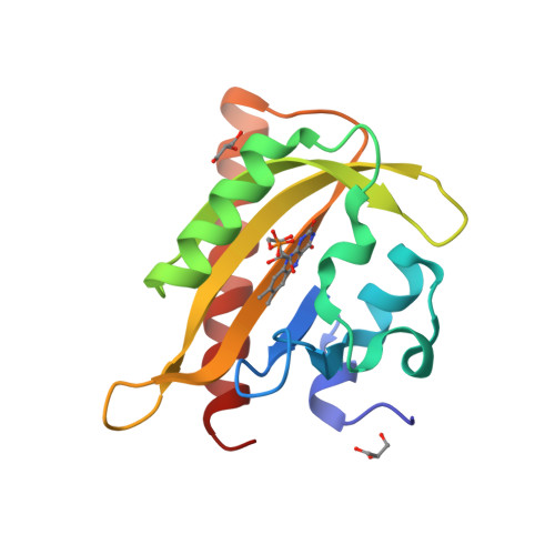
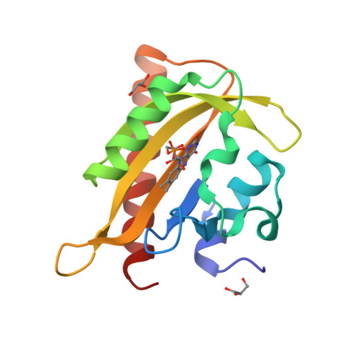
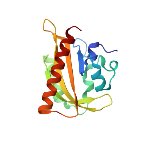
N- and C-Terminal Flanking Regions Modulate Light-Induced Signal Transduction in the Lov2 Domain of the Blue Light Sensor Phototropin 1 from Avena Sativa.

Halavaty, A.S.Moffat, K.

(2007) Biochemistry 46: 14001

  • DOI: https://doi.org/10.1021/bi701543e
  • Primary Citation of Related Structures:  
    2V0U, 2V0W, 2V1A, 2V1B

  • PubMed Abstract: 

    Light sensing by photoreceptors controls phototropism, chloroplast movement, stomatal opening, and leaf expansion in plants. Understanding the molecular mechanism by which these processes are regulated requires a quantitative description of photoreceptor dynamics. We focus on a light-driven signal transduction mechanism in the LOV2 domain (LOV, light, oxygen, voltage) of the blue light photoreceptor phototropin 1 from Avena sativa (oat). High-resolution crystal structures of the dark and light states of an oat LOV2 construct including residues Leu404 through Leu546 (LOV2 (404-546)) have been determined at 105 and 293 K. In all four structures, LOV2 (404-546) exhibits the typical Per-ARNT-Sim (PAS) fold, flanked by an additional conserved N-terminal turn-helix-turn motif and a C-terminal flanking region containing an amphipathic Jalpha helix. These regions dock on the LOV2 core domain and bury several hydrophobic residues of the central beta-sheet of the core domain that would otherwise be exposed to solvent. Light structures of LOV2 (404-546) reveal that formation of the covalent bond between Cys450 and the C4a atom of the flavin mononucleotide (FMN) results in local rearrangement of the hydrogen-bonding network in the FMN binding pocket. These rearrangements are associated with disruption of the Asn414-Asp515 hydrogen bond on the surface of the protein and displacement of the N- and C-terminal flanking regions of LOV2 (404-546), both of which constitute a structural signal.


  • Organizational Affiliation

    Department of Biochemistry and Molecular Biology, The University of Chicago, 929 East 57th Street, Chicago, Illinois 60637, USA.


Macromolecules
Find similar proteins by:  (by identity cutoff)  |  3D Structure
Entity ID: 1
MoleculeChains Sequence LengthOrganismDetailsImage
NPH1-1144Avena sativaMutation(s): 0 
EC: 2.7.11.1
UniProt
Find proteins for O49003 (Avena sativa)
Explore O49003 
Go to UniProtKB:  O49003
Entity Groups  
Sequence Clusters30% Identity50% Identity70% Identity90% Identity95% Identity100% Identity
UniProt GroupO49003
Sequence Annotations
Expand
  • Reference Sequence
Experimental Data & Validation

Experimental Data

  • Method: X-RAY DIFFRACTION
  • Resolution: 1.65 Å
  • R-Value Free:  0.195 (Depositor), 0.210 (DCC) 
  • R-Value Work:  0.156 (Depositor), 0.170 (DCC) 
  • R-Value Observed: 0.158 (Depositor) 
Space Group: P 21 21 21
Unit Cell:
Length ( Å )Angle ( ˚ )
a = 35.706α = 90
b = 57.155β = 90
c = 67.395γ = 90
Software Package:
Software NamePurpose
REFMACrefinement
HKL-2000data reduction
HKL-2000data scaling
AMoREphasing

Structure Validation

View Full Validation Report


Ligand Structure Quality Assessment 

Created with Raphaël 2.3.0Worse 01 BetterLigand structure goodness of fit to experimental dataBest fitted FMNClick on this verticalbar to view details

Entry History 

Deposition Data

Revision History  (Full details and data files)

  • Version 1.0: 2007-12-11
    Type: Initial release
  • Version 1.1: 2011-07-13
    Changes: Advisory, Version format compliance
  • Version 1.2: 2019-10-16
    Changes: Data collection, Experimental preparation, Other
  • Version 1.3: 2023-12-13
    Changes: Data collection, Database references, Refinement description