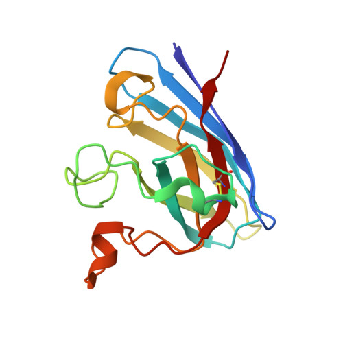
2WYT

1.0 A resolution structure of L38V SOD1 mutant


Experimental Data Snapshot

  • Method: X-RAY DIFFRACTION
  • Resolution: 1.00 Å
  • R-Value Free: 0.147 
  • R-Value Work: 0.124 
  • R-Value Observed: 0.125 

Starting Model: experimental
View more details

wwPDB Validation   3D Report Full Report


This is version 2.1 of the entry. See complete history


Literature

Structural Discovery of Small Molecule Binding Sites in Cu-Zn Human Superoxide Dismutase Familial Amyotrophic Lateral Sclerosis Mutants Provides Insights for Lead Optimization.

Antonyuk, S.V.Strange, R.W.Hasnain, S.S.

(2010) J Med Chem 53: 1402

  • DOI: https://doi.org/10.1021/jm9017948
  • Primary Citation of Related Structures:  
    2WYT, 2WYZ, 2WZ0, 2WZ5, 2WZ6

  • PubMed Abstract: 

    Dominant inheritance of point mutations in CuZn superoxide dismutase (SOD1) is the best characterized subset of familial amyotrophic lateral sclerosis (FALS) and accounts for some 20% of the known familial cases. We report the discovery and visualization via cocrystallography of two ligand-binding pockets in human SOD1 and its pathogenic mutants that have opened up the real possibility of undertaking lead compound discovery using a fragment-based approach for therapeutic purposes for SOD1 associated motor neuron disease.


  • Organizational Affiliation

    Molecular Biophysics Group, School of Biological Sciences, University of Liverpool, Liverpool L69 7ZB, UK.


Macromolecules
Find similar proteins by:  (by identity cutoff)  |  3D Structure
Entity ID: 1
MoleculeChains Sequence LengthOrganismDetailsImage
SUPEROXIDE DISMUTASE [CU-ZN]A,
B [auth F]
153Homo sapiensMutation(s): 1 
EC: 1.15.1.1
UniProt & NIH Common Fund Data Resources
Find proteins for P00441 (Homo sapiens)
Explore P00441 
Go to UniProtKB:  P00441
PHAROS:  P00441
GTEx:  ENSG00000142168 
Entity Groups  
Sequence Clusters30% Identity50% Identity70% Identity90% Identity95% Identity100% Identity
UniProt GroupP00441
Sequence Annotations
Expand
  • Reference Sequence
Small Molecules
Ligands 5 Unique
IDChains Name / Formula / InChI Key2D Diagram3D Interactions
SO4
Query on SO4

Download Ideal Coordinates CCD File 
F [auth A]
G [auth A]
I [auth A]
K [auth F]
L [auth F]
F [auth A],
G [auth A],
I [auth A],
K [auth F],
L [auth F],
P [auth F]
SULFATE ION
O4 S
QAOWNCQODCNURD-UHFFFAOYSA-L
ZN
Query on ZN

Download Ideal Coordinates CCD File 
D [auth A],
E [auth A],
N [auth F],
O [auth F]
ZINC ION
Zn
PTFCDOFLOPIGGS-UHFFFAOYSA-N
CU
Query on CU

Download Ideal Coordinates CCD File 
C [auth A],
M [auth F]
COPPER (II) ION
Cu
JPVYNHNXODAKFH-UHFFFAOYSA-N
ACT
Query on ACT

Download Ideal Coordinates CCD File 
H [auth A]ACETATE ION
C2 H3 O2
QTBSBXVTEAMEQO-UHFFFAOYSA-M
CL
Query on CL

Download Ideal Coordinates CCD File 
J [auth A],
Q [auth F]
CHLORIDE ION
Cl
VEXZGXHMUGYJMC-UHFFFAOYSA-M
Experimental Data & Validation

Experimental Data

  • Method: X-RAY DIFFRACTION
  • Resolution: 1.00 Å
  • R-Value Free: 0.147 
  • R-Value Work: 0.124 
  • R-Value Observed: 0.125 
  • Space Group: P 1 21 1
Unit Cell:
Length ( Å )Angle ( ˚ )
a = 38.69α = 90
b = 67.345β = 106.57
c = 52.577γ = 90
Software Package:
Software NamePurpose
REFMACrefinement
HKL-2000data reduction
HKL-2000data scaling
MOLREPphasing

Structure Validation

View Full Validation Report


Entry History 

Deposition Data

Revision History  (Full details and data files)

  • Version 1.0: 2010-10-27
    Type: Initial release
  • Version 1.1: 2011-05-08
    Changes: Version format compliance
  • Version 1.2: 2011-07-13
    Changes: Version format compliance
  • Version 1.3: 2017-07-12
    Changes: Derived calculations
  • Version 2.0: 2023-12-20
    Changes: Advisory, Atomic model, Data collection, Database references, Derived calculations, Other, Refinement description
  • Version 2.1: 2024-11-13
    Changes: Structure summary