3A3Y | pdb_00003a3y

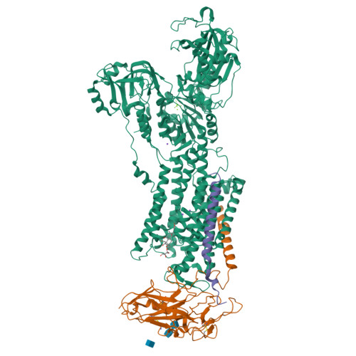
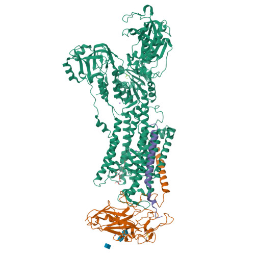
Crystal structure of the sodium-potassium pump with bound potassium and ouabain


Experimental Data Snapshot

  • Method: X-RAY DIFFRACTION
  • Resolution: 2.80 Å
  • R-Value Free: 
    0.293 (Depositor), 0.289 (DCC) 
  • R-Value Work: 
    0.247 (Depositor), 0.245 (DCC) 
  • R-Value Observed: 
    0.249 (Depositor) 

wwPDB Validation   3D Report Full Report


Ligand Structure Quality Assessment 

Created with Raphaël 2.3.0Worse 01 BetterLigand structure goodness of fit to experimental dataBest fitted OBNClick on this verticalbar to view detailsBest fitted CLRClick on this verticalbar to view detailsBest fitted NAGClick on this verticalbar to view details

This is version 1.3 of the entry. See complete history


Literature

Crystal structure of the sodium-potassium pump (Na+,K+-ATPase) with bound potassium and ouabain.

Ogawa, H.Shinoda, T.Cornelius, F.Toyoshima, C.

(2009) Proc Natl Acad Sci U S A 106: 13742-13747

  • DOI: https://doi.org/10.1073/pnas.0907054106
  • Primary Citation of Related Structures:  
    3A3Y

  • PubMed Abstract: 

    The sodium-potassium pump (Na(+),K(+)-ATPase) is responsible for establishing Na(+) and K(+) concentration gradients across the plasma membrane and therefore plays an essential role in, for instance, generating action potentials. Cardiac glycosides, prescribed for congestive heart failure for more than 2 centuries, are efficient inhibitors of this ATPase. Here we describe a crystal structure of Na(+),K(+)-ATPase with bound ouabain, a representative cardiac glycoside, at 2.8 A resolution in a state analogous to E2.2K(+).Pi. Ouabain is deeply inserted into the transmembrane domain with the lactone ring very close to the bound K(+), in marked contrast to previous models. Due to antagonism between ouabain and K(+), the structure represents a low-affinity ouabain-bound state. Yet, most of the mutagenesis data obtained with the high-affinity state are readily explained by the present crystal structure, indicating that the binding site for ouabain is essentially the same. According to a homology model for the high affinity state, it is a closure of the binding cavity that confers a high affinity.


  • Organizational Affiliation

    Institute of Molecular and Cellular Biosciences, The University of Tokyo, Bunkyo-ku, Tokyo 113-0032, Japan.


Macromolecules
Find similar proteins by:  (by identity cutoff)  |  3D Structure
Entity ID: 1
MoleculeChains Sequence LengthOrganismDetailsImage
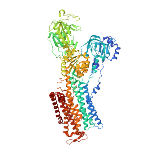
Na, K-ATPase alpha subunit1,028Squalus acanthiasMutation(s): 0 
Membrane Entity: Yes 
UniProt
Find proteins for Q4H132 (Squalus acanthias)
Explore Q4H132 
Go to UniProtKB:  Q4H132
Entity Groups  
Sequence Clusters30% Identity50% Identity70% Identity90% Identity95% Identity100% Identity
UniProt GroupQ4H132
Sequence Annotations
Expand
  • Reference Sequence
Find similar proteins by:  (by identity cutoff)  |  3D Structure
Entity ID: 2
MoleculeChains Sequence LengthOrganismDetailsImage
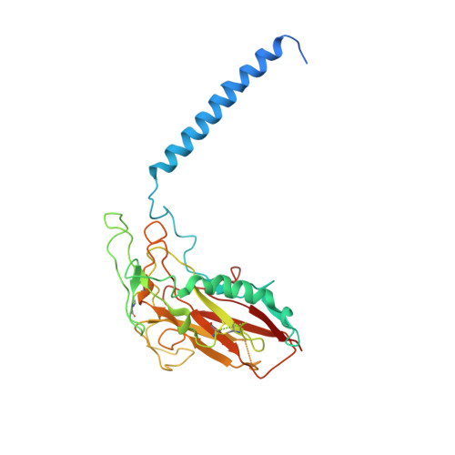
NA+,K+-ATPASE BETA SUBUNIT305Squalus acanthiasMutation(s): 0 
Membrane Entity: Yes 
UniProt
Find proteins for C4IX13 (Squalus acanthias)
Explore C4IX13 
Go to UniProtKB:  C4IX13
Entity Groups  
Sequence Clusters30% Identity50% Identity70% Identity90% Identity95% Identity100% Identity
UniProt GroupC4IX13
Sequence Annotations
Expand
  • Reference Sequence
Find similar proteins by:  (by identity cutoff)  |  3D Structure
Entity ID: 3
MoleculeChains Sequence LengthOrganismDetailsImage
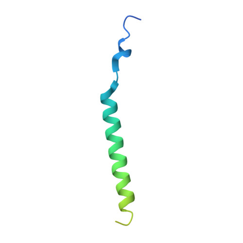
Phospholemman-like proteinC [auth G]74Squalus acanthiasMutation(s): 0 
Membrane Entity: Yes 
UniProt
Find proteins for Q70Q12 (Squalus acanthias)
Explore Q70Q12 
Go to UniProtKB:  Q70Q12
Entity Groups  
Sequence Clusters30% Identity50% Identity70% Identity90% Identity95% Identity100% Identity
UniProt GroupQ70Q12
Sequence Annotations
Expand
  • Reference Sequence
Small Molecules
Ligands 6 Unique
IDChains Name / Formula / InChI Key2D Diagram3D Interactions
OBN
Query on OBN

Download Ideal Coordinates CCD File 
I [auth A]OUABAIN
C29 H44 O12
LPMXVESGRSUGHW-HBYQJFLCSA-N
CLR
Query on CLR

Download Ideal Coordinates CCD File 
J [auth B]CHOLESTEROL
C27 H46 O
HVYWMOMLDIMFJA-DPAQBDIFSA-N
NAG
Query on NAG

Download Ideal Coordinates CCD File 
K [auth B],
L [auth B],
M [auth B]
2-acetamido-2-deoxy-beta-D-glucopyranose
C8 H15 N O6
OVRNDRQMDRJTHS-FMDGEEDCSA-N
MF4
Query on MF4

Download Ideal Coordinates CCD File 
D [auth A]TETRAFLUOROMAGNESATE(2-)
F4 Mg
XVYWAXYEHHUKQW-UHFFFAOYSA-J
K
Query on K

Download Ideal Coordinates CCD File 
F [auth A],
G [auth A],
H [auth A]
POTASSIUM ION
K
NPYPAHLBTDXSSS-UHFFFAOYSA-N
MG
Query on MG

Download Ideal Coordinates CCD File 
E [auth A]MAGNESIUM ION
Mg
JLVVSXFLKOJNIY-UHFFFAOYSA-N
Binding Affinity Annotations 
IDSourceBinding Affinity
OBN BindingDB:  3A3Y Ki: 97 (nM) from 1 assay(s)
IC50: min: 31, max: 4300 (nM) from 2 assay(s)
Experimental Data & Validation

Experimental Data

  • Method: X-RAY DIFFRACTION
  • Resolution: 2.80 Å
  • R-Value Free:  0.293 (Depositor), 0.289 (DCC) 
  • R-Value Work:  0.247 (Depositor), 0.245 (DCC) 
  • R-Value Observed: 0.249 (Depositor) 
Space Group: C 1 2 1
Unit Cell:
Length ( Å )Angle ( ˚ )
a = 222.884α = 90
b = 50.72β = 104.63
c = 163.342γ = 90
Software Package:
Software NamePurpose
REFMACrefinement
DENZOdata reduction
SCALEPACKdata scaling
CNSphasing

Structure Validation

View Full Validation Report


Ligand Structure Quality Assessment 

Created with Raphaël 2.3.0Worse 01 BetterLigand structure goodness of fit to experimental dataBest fitted OBNClick on this verticalbar to view detailsBest fitted CLRClick on this verticalbar to view detailsBest fitted NAGClick on this verticalbar to view details

Entry History 

Deposition Data

Revision History  (Full details and data files)

  • Version 1.0: 2009-09-08
    Type: Initial release
  • Version 1.1: 2011-07-13
    Changes: Advisory, Version format compliance
  • Version 1.2: 2020-07-29
    Type: Remediation
    Reason: Carbohydrate remediation
    Changes: Data collection, Derived calculations, Structure summary
  • Version 1.3: 2024-11-13
    Changes: Data collection, Database references, Structure summary