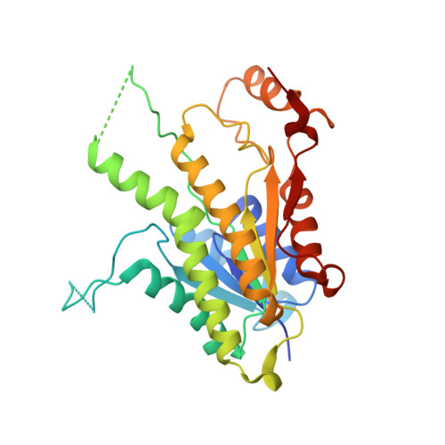
3H4V

Selective screening and design to identify inhibitors of leishmania major pteridine reductase 1


Experimental Data Snapshot

  • Method: X-RAY DIFFRACTION
  • Resolution: 2.40 Å
  • R-Value Free: 0.356 
  • R-Value Work: 0.276 
  • R-Value Observed: 0.280 

Starting Model: experimental
View more details

wwPDB Validation   3D Report Full Report


Ligand Structure Quality Assessment 


This is version 1.3 of the entry. See complete history


Literature

Discovery of potent pteridine reductase inhibitors to guide antiparasite drug development

Cavazzuti, A.Paglietti, G.Hunter, W.N.Gamarro, F.Piras, S.Loriga, M.Allecca, S.Corona, P.McLuskey, K.Tulloch, L.Gibellini, F.Ferrari, S.Costi, M.P.

(2008) Proc Natl Acad Sci U S A 105: 1448-1453

  • DOI: https://doi.org/10.1073/pnas.0704384105
  • Primary Citation of Related Structures:  
    2QHX, 3H4V

  • PubMed Abstract: 

    Pteridine reductase (PTR1) is essential for salvage of pterins by parasitic trypanosomatids and is a target for the development of improved therapies. To identify inhibitors of Leishmania major and Trypanosoma cruzi PTR1, we combined a rapid-screening strategy using a folate-based library with structure-based design. Assays were carried out against folate-dependent enzymes including PTR1, dihydrofolate reductase (DHFR), and thymidylate synthase. Affinity profiling determined selectivity and specificity of a series of quinoxaline and 2,4-diaminopteridine derivatives, and nine compounds showed greater activity against parasite enzymes compared with human enzymes. Compound 6a displayed a K(i) of 100 nM toward LmPTR1, and the crystal structure of the LmPTR1:NADPH:6a ternary complex revealed a substrate-like binding mode distinct from that previously observed for similar compounds. A second round of design, synthesis, and assay produced a compound (6b) with a significantly improved K(i) (37 nM) against LmPTR1, and the structure of this complex was also determined. Biological evaluation of selected inhibitors was performed against the extracellular forms of T. cruzi and L. major, both wild-type and overexpressing PTR1 lines, as a model for PTR1-driven antifolate drug resistance and the intracellular form of T. cruzi. An additive profile was observed when PTR1 inhibitors were used in combination with known DHFR inhibitors, and a reduction in toxicity of treatment was observed with respect to administration of a DHFR inhibitor alone. The successful combination of antifolates targeting two enzymes indicates high potential for such an approach in the development of previously undescribed antiparasitic drugs.


  • Organizational Affiliation

    Dipartimento di Scienze Farmaceutiche, Università di Modena e Reggio Emilia, Via Campi 183, 41100 Modena, Italy.


Macromolecules
Find similar proteins by:  (by identity cutoff)  |  3D Structure
Entity ID: 1
MoleculeChains Sequence LengthOrganismDetailsImage
Pteridine reductase 1
A, B, C, D, E
A, B, C, D, E, F, G, H
288Leishmania majorMutation(s): 0 
Gene Names: PTR1HMTXR
EC: 1.5.1.33
UniProt
Find proteins for Q01782 (Leishmania major)
Explore Q01782 
Go to UniProtKB:  Q01782
Entity Groups  
Sequence Clusters30% Identity50% Identity70% Identity90% Identity95% Identity100% Identity
UniProt GroupQ01782
Sequence Annotations
Expand
  • Reference Sequence
Small Molecules
Ligands 2 Unique
IDChains Name / Formula / InChI Key2D Diagram3D Interactions
NAP
Query on NAP

Download Ideal Coordinates CCD File 
I [auth A]
K [auth B]
M [auth C]
O [auth D]
Q [auth E]
I [auth A],
K [auth B],
M [auth C],
O [auth D],
Q [auth E],
S [auth F],
U [auth G],
W [auth H]
NADP NICOTINAMIDE-ADENINE-DINUCLEOTIDE PHOSPHATE
C21 H28 N7 O17 P3
XJLXINKUBYWONI-NNYOXOHSSA-N
DVP
Query on DVP

Download Ideal Coordinates CCD File 
J [auth A]
L [auth B]
N [auth C]
P [auth D]
R [auth E]
J [auth A],
L [auth B],
N [auth C],
P [auth D],
R [auth E],
T [auth F],
V [auth G],
X [auth H]
METHYL 1-(4-{[(2,4-DIAMINOPTERIDIN-6-YL)METHYL]AMINO}BENZOYL)PIPERIDINE-4-CARBOXYLATE
C21 H24 N8 O3
NYNAFINLHQEHKU-UHFFFAOYSA-N
Binding Affinity Annotations 
IDSourceBinding Affinity
DVP BindingDB:  3H4V Ki: 100 (nM) from 1 assay(s)
IC50: 1690 (nM) from 1 assay(s)
Experimental Data & Validation

Experimental Data

  • Method: X-RAY DIFFRACTION
  • Resolution: 2.40 Å
  • R-Value Free: 0.356 
  • R-Value Work: 0.276 
  • R-Value Observed: 0.280 
  • Space Group: P 1 21 1
Unit Cell:
Length ( Å )Angle ( ˚ )
a = 95.206α = 90
b = 103.492β = 108.24
c = 146.476γ = 90
Software Package:
Software NamePurpose
AMoREphasing
REFMACrefinement
HKL-2000data reduction
DENZOdata reduction
SCALEPACKdata scaling

Structure Validation

View Full Validation Report


Ligand Structure Quality Assessment 


Entry History 

Deposition Data

Revision History  (Full details and data files)

  • Version 1.0: 2009-05-05
    Type: Initial release
  • Version 1.1: 2011-07-13
    Changes: Version format compliance
  • Version 1.2: 2017-11-01
    Changes: Refinement description
  • Version 1.3: 2023-11-01
    Changes: Data collection, Database references, Derived calculations, Refinement description