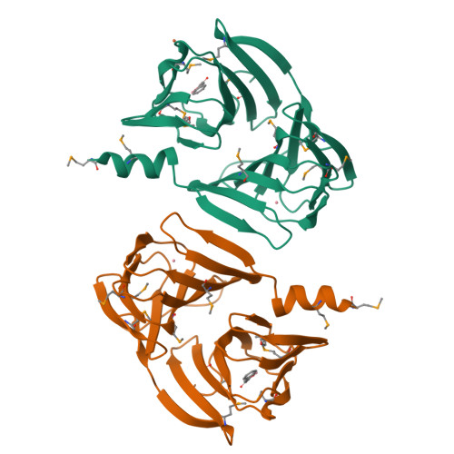
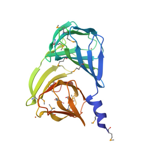
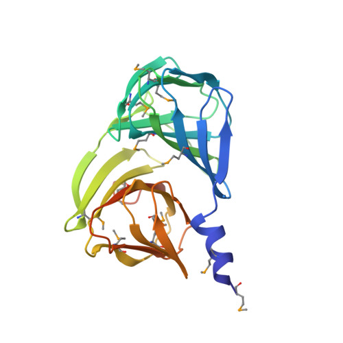
3H9A

Crystal structure of BacB, an enzyme involved in Bacilysin synthesis, in triclinic form


Experimental Data Snapshot

  • Method: X-RAY DIFFRACTION
  • Resolution: 2.04 Å
  • R-Value Free: 0.262 
  • R-Value Work: 0.206 
  • R-Value Observed: 0.209 

Starting Model: experimental
View more details

wwPDB Validation   3D Report Full Report


Ligand Structure Quality Assessment 


This is version 1.4 of the entry. See complete history


Literature

Analysis of multiple crystal forms of Bacillus subtilis BacB suggests a role for a metal ion as a nucleant for crystallization

Rajavel, M.Gopal, B.

(2010) Acta Crystallogr D Biol Crystallogr 66: 635-639

  • DOI: https://doi.org/10.1107/S0907444910006682
  • Primary Citation of Related Structures:  
    3H9A

  • PubMed Abstract: 

    Bacillus subtilis BacB is an oxidase that is involved in the production of the antibiotic bacilysin. This protein contains two double-stranded beta-helix (cupin) domains fused in a compact arrangement. BacB crystallizes in three crystal forms under similar crystallization conditions. An interesting observation was that a slight perturbation of the crystallization droplet resulted in the nucleation of a different crystal form. An X-ray absorption scan of BacB suggested the presence of cobalt and iron in the crystal. Here, a comparative analysis of the different crystal forms of BacB is presented in an effort to identify the basis for the different lattices. It is noted that metal ions mediating interactions across the asymmetric unit dominate the different packing arrangements. Furthermore, a normalized B-factor analysis of all the crystal structures suggests that the solvent-exposed metal ions decrease the flexibility of a loop segment, perhaps influencing the choice of crystal form. The residues coordinating the surface metal ion are similar in the triclinic and monoclinic crystal forms. The coordinating ligands for the corresponding metal ion in the tetragonal crystal form are different, leading to a tighter packing arrangement. Although BacB is a monomer in solution, a dimer of BacB serves as a template on which higher order symmetrical arrangements are formed. The different crystal forms of BacB thus provide experimental evidence for metal-ion-mediated lattice formation and crystal packing.


  • Organizational Affiliation

    Molecular Biophysics Unit, Indian Institute of Science, Bangalore 560 012, India.


Macromolecules
Find similar proteins by:  (by identity cutoff)  |  3D Structure
Entity ID: 1
MoleculeChains Sequence LengthOrganismDetailsImage
Bacilysin biosynthesis protein bacB
A, B
243Bacillus subtilisMutation(s): 0 
Gene Names: ywfCbacB
EC: 5.3.3.19
UniProt
Find proteins for P39639 (Bacillus subtilis (strain 168))
Explore P39639 
Go to UniProtKB:  P39639
Entity Groups  
Sequence Clusters30% Identity50% Identity70% Identity90% Identity95% Identity100% Identity
UniProt GroupP39639
Sequence Annotations
Expand
  • Reference Sequence
Small Molecules
Ligands 3 Unique
IDChains Name / Formula / InChI Key2D Diagram3D Interactions
PPY
Query on PPY

Download Ideal Coordinates CCD File 
E [auth A],
I [auth B]
3-PHENYLPYRUVIC ACID
C9 H8 O3
BTNMPGBKDVTSJY-UHFFFAOYSA-N
CO
Query on CO

Download Ideal Coordinates CCD File 
C [auth A],
D [auth A],
G [auth B],
H [auth B]
COBALT (II) ION
Co
XLJKHNWPARRRJB-UHFFFAOYSA-N
FE
Query on FE

Download Ideal Coordinates CCD File 
F [auth A]FE (III) ION
Fe
VTLYFUHAOXGGBS-UHFFFAOYSA-N
Modified Residues  1 Unique
IDChains TypeFormula2D DiagramParent
MSE
Query on MSE
A, B
L-PEPTIDE LINKINGC5 H11 N O2 SeMET
Experimental Data & Validation

Experimental Data

  • Method: X-RAY DIFFRACTION
  • Resolution: 2.04 Å
  • R-Value Free: 0.262 
  • R-Value Work: 0.206 
  • R-Value Observed: 0.209 
  • Space Group: P 1
Unit Cell:
Length ( Å )Angle ( ˚ )
a = 46.252α = 89.05
b = 47.198β = 77.27
c = 62.906γ = 82.28
Software Package:
Software NamePurpose
MAR345dtbdata collection
PHASESphasing
REFMACrefinement
MOSFLMdata reduction
SCALAdata scaling

Structure Validation

View Full Validation Report


Ligand Structure Quality Assessment 


Entry History 

Deposition Data

Revision History  (Full details and data files)

  • Version 1.0: 2010-03-02
    Type: Initial release
  • Version 1.1: 2011-07-13
    Changes: Advisory, Version format compliance
  • Version 1.2: 2023-11-01
    Changes: Data collection, Database references, Derived calculations, Refinement description
  • Version 1.3: 2023-11-22
    Changes: Data collection
  • Version 1.4: 2024-10-09
    Changes: Structure summary