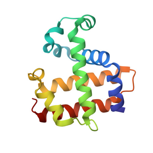
3LB3

Two-site competitive inhibition in dehaloperoxidase-hemoglobin


Experimental Data Snapshot

  • Method: X-RAY DIFFRACTION
  • Resolution: 1.85 Å
  • R-Value Free: 0.243 
  • R-Value Work: 0.187 
  • R-Value Observed: 0.190 

Starting Model: experimental
View more details

wwPDB Validation   3D Report Full Report


Ligand Structure Quality Assessment 


This is version 1.2 of the entry. See complete history


Literature

Internal binding of halogenated phenols in dehaloperoxidase-hemoglobin inhibits peroxidase function.

Thompson, M.K.Davis, M.F.de Serrano, V.Nicoletti, F.P.Howes, B.D.Smulevich, G.Franzen, S.

(2010) Biophys J 99: 1586-1595

  • DOI: https://doi.org/10.1016/j.bpj.2010.05.041
  • Primary Citation of Related Structures:  
    3LB1, 3LB2, 3LB3, 3LB4

  • PubMed Abstract: 

    Dehaloperoxidase (DHP) from the annelid Amphitrite ornata is a catalytically active hemoglobin-peroxidase that possesses a unique internal binding cavity in the distal pocket above the heme. The previously published crystal structure of DHP shows 4-iodophenol bound internally. This led to the proposal that the internal binding site is the active site for phenol oxidation. However, the native substrate for DHP is 2,4,6-tribromophenol, and all attempts to bind 2,4,6-tribromophenol in the internal site under physiological conditions have failed. Herein, we show that the binding of 4-halophenols in the internal pocket inhibits enzymatic function. Furthermore, we demonstrate that DHP has a unique two-site competitive binding mechanism in which the internal and external binding sites communicate through two conformations of the distal histidine of the enzyme, resulting in nonclassical competitive inhibition. The same distal histidine conformations involved in DHP function regulate oxygen binding and release during transport and storage by hemoglobins and myoglobins. This work provides further support for the hypothesis that DHP possesses an external binding site for substrate oxidation, as is typical for the peroxidase family of enzymes.


  • Organizational Affiliation

    Department of Chemistry, North Carolina State University, Raleigh, North Carolina, USA.


Macromolecules
Find similar proteins by:  (by identity cutoff)  |  3D Structure
Entity ID: 1
MoleculeChains Sequence LengthOrganismDetailsImage
Dehaloperoxidase A
A, B
137Amphitrite ornataMutation(s): 0 
Gene Names: DHPA
UniProt
Find proteins for Q9NAV8 (Amphitrite ornata)
Explore Q9NAV8 
Go to UniProtKB:  Q9NAV8
Entity Groups  
Sequence Clusters30% Identity50% Identity70% Identity90% Identity95% Identity100% Identity
UniProt GroupQ9NAV8
Sequence Annotations
Expand
  • Reference Sequence
Small Molecules
Ligands 3 Unique
IDChains Name / Formula / InChI Key2D Diagram3D Interactions
HEM
Query on HEM

Download Ideal Coordinates CCD File 
C [auth A],
H [auth B]
PROTOPORPHYRIN IX CONTAINING FE
C34 H32 Fe N4 O4
KABFMIBPWCXCRK-RGGAHWMASA-L
4CH
Query on 4CH

Download Ideal Coordinates CCD File 
D [auth A],
I [auth B]
4-chlorophenol
C6 H5 Cl O
WXNZTHHGJRFXKQ-UHFFFAOYSA-N
SO4
Query on SO4

Download Ideal Coordinates CCD File 
E [auth A],
F [auth A],
G [auth B]
SULFATE ION
O4 S
QAOWNCQODCNURD-UHFFFAOYSA-L
Experimental Data & Validation

Experimental Data

  • Method: X-RAY DIFFRACTION
  • Resolution: 1.85 Å
  • R-Value Free: 0.243 
  • R-Value Work: 0.187 
  • R-Value Observed: 0.190 
  • Space Group: P 21 21 21
Unit Cell:
Length ( Å )Angle ( ˚ )
a = 58.201α = 90
b = 67.21β = 90
c = 68.724γ = 90
Software Package:
Software NamePurpose
HKL-2000data collection
PHASERphasing
REFMACrefinement
DENZOdata reduction
SCALEPACKdata scaling

Structure Validation

View Full Validation Report


Ligand Structure Quality Assessment 


Entry History 

Deposition Data

Revision History  (Full details and data files)

  • Version 1.0: 2010-11-24
    Type: Initial release
  • Version 1.1: 2011-07-13
    Changes: Version format compliance
  • Version 1.2: 2023-09-06
    Changes: Data collection, Database references, Derived calculations, Refinement description