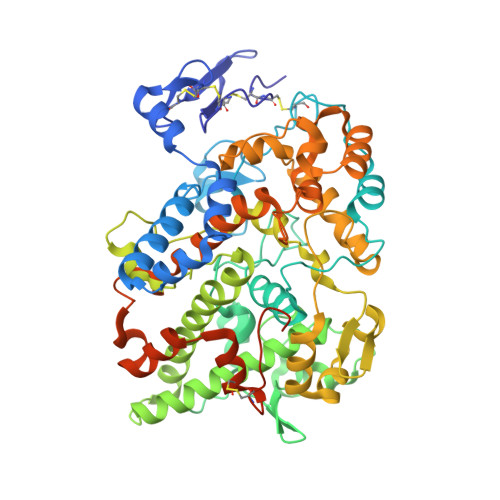
3LN0

Structure of compound 5c-S bound at the active site of COX-2


Experimental Data Snapshot

  • Method: X-RAY DIFFRACTION
  • Resolution: 2.20 Å
  • R-Value Free: 0.238 
  • R-Value Work: 0.209 
  • R-Value Observed: 0.212 

wwPDB Validation   3D Report Full Report


Ligand Structure Quality Assessment 


This is version 2.0 of the entry. See complete history


Literature

The novel benzopyran class of selective cyclooxygenase-2 inhibitors. Part 2: The second clinical candidate having a shorter and favorable human half-life.

Wang, J.L.Limburg, D.Graneto, M.J.Springer, J.Hamper, J.R.Liao, S.Pawlitz, J.L.Kurumbail, R.G.Maziasz, T.Talley, J.J.Kiefer, J.R.Carter, J.

(2010) Bioorg Med Chem Lett 20: 7159-7163

  • DOI: https://doi.org/10.1016/j.bmcl.2010.07.054
  • Primary Citation of Related Structures:  
    3LN0, 3LN1, 3MQE, 3NTG

  • PubMed Abstract: 

    In this Letter, we provide the structure-activity relationships, optimization of design, testing criteria, and human half-life data for a series of selective COX-2 inhibitors. During the course of our structure-based drug design efforts, we discovered two distinct binding modes within the COX-2 active site for differently substituted members of this class. The challenge of a undesirably long human half-life for the first clinical candidate 1t(1/2)=360 h was addressed by multiple strategies, leading to the discovery of 29b-(S) (SC-75416) with t(1/2)=34 h.


  • Organizational Affiliation

    Pfizer Global Research and Development, Chesterfield, MO 63017, USA. jl47wang@yahoo.com


Macromolecules
Find similar proteins by:  (by identity cutoff)  |  3D Structure
Entity ID: 1
MoleculeChains Sequence LengthOrganismDetailsImage
Prostaglandin G/H synthase 2
A, B, C, D
587Mus musculusMutation(s): 0 
Gene Names: Cox-2Cox2Pghs-bprostaglandin H2 synthase 2Ptgs2Tis10
EC: 1.14.99.1
UniProt
Find proteins for Q05769 (Mus musculus)
Go to UniProtKB:  Q05769
Glycosylation
Glycosylation Sites: 3Go to GlyGen: Q05769-1
Sequence Annotations
Expand
  • Reference Sequence
Oligosaccharides

Help

Entity ID: 2
MoleculeChains Length2D Diagram Glycosylation3D Interactions
2-acetamido-2-deoxy-beta-D-glucopyranose-(1-4)-2-acetamido-2-deoxy-beta-D-glucopyranose-(1-4)-2-acetamido-2-deoxy-beta-D-glucopyranose
E, F, G, H
3N-Glycosylation
Glycosylation Resources
GlyTouCan:  G47362BJ
GlyCosmos:  G47362BJ
GlyGen:  G47362BJ
Small Molecules
Ligands 4 Unique
IDChains Name / Formula / InChI Key2D Diagram3D Interactions
HEM
Query on HEM

Download Ideal Coordinates CCD File 
I [auth A],
N [auth B],
R [auth C],
V [auth D]
PROTOPORPHYRIN IX CONTAINING FE
C34 H32 Fe N4 O4
KABFMIBPWCXCRK-RGGAHWMASA-L
52B
Query on 52B

Download Ideal Coordinates CCD File 
L [auth A],
Q [auth B],
U [auth C],
Z [auth D]
(2S)-6,8-dichloro-2-(trifluoromethyl)-2H-chromene-3-carboxylic acid
C11 H5 Cl2 F3 O3
ZFKBWSREWJOSSJ-VIFPVBQESA-N
BOG
Query on BOG

Download Ideal Coordinates CCD File 
M [auth A],
Y [auth D]
octyl beta-D-glucopyranoside
C14 H28 O6
HEGSGKPQLMEBJL-RKQHYHRCSA-N
NAG
Query on NAG

Download Ideal Coordinates CCD File 
J [auth A]
K [auth A]
O [auth B]
P [auth B]
S [auth C]
J [auth A],
K [auth A],
O [auth B],
P [auth B],
S [auth C],
T [auth C],
W [auth D],
X [auth D]
2-acetamido-2-deoxy-beta-D-glucopyranose
C8 H15 N O6
OVRNDRQMDRJTHS-FMDGEEDCSA-N
Binding Affinity Annotations 
IDSourceBinding Affinity
52B BindingDB:  3LN0 IC50: min: 9.8, max: 300 (nM) from 4 assay(s)
Biologically Interesting Molecules (External Reference) 1 Unique
Experimental Data & Validation

Experimental Data

  • Method: X-RAY DIFFRACTION
  • Resolution: 2.20 Å
  • R-Value Free: 0.238 
  • R-Value Work: 0.209 
  • R-Value Observed: 0.212 
  • Space Group: P 21 21 2
Unit Cell:
Length ( Å )Angle ( ˚ )
a = 181.015α = 90
b = 135.201β = 90
c = 124.291γ = 90
Software Package:
Software NamePurpose
MAR345dtbdata collection
PHASERphasing
REFMACrefinement
HKL-2000data reduction
HKL-2000data scaling

Structure Validation

View Full Validation Report


Ligand Structure Quality Assessment 


Entry History 

Deposition Data

Revision History  (Full details and data files)

  • Version 1.0: 2010-10-27
    Type: Initial release
  • Version 1.1: 2011-07-13
    Changes: Version format compliance
  • Version 2.0: 2020-07-29
    Type: Remediation
    Reason: Carbohydrate remediation
    Changes: Atomic model, Data collection, Derived calculations, Structure summary