3MOO | pdb_00003moo

Crystal structure of the HmuO, heme oxygenase from Corynebacterium diphtheriae, in complex with azide-bound verdoheme


Experimental Data Snapshot

  • Method: X-RAY DIFFRACTION
  • Resolution: 1.71 Å
  • R-Value Free: 
    0.213 (Depositor), 0.210 (DCC) 
  • R-Value Work: 
    0.178 (Depositor), 0.180 (DCC) 
  • R-Value Observed: 
    0.182 (Depositor) 

Starting Model: experimental
View more details

wwPDB Validation   3D Report Full Report


Ligand Structure Quality Assessment 

Created with Raphaël 2.3.0Worse 01 BetterLigand structure goodness of fit to experimental dataBest fitted VEAClick on this verticalbar to view details

This is version 2.1 of the entry. See complete history


Literature

Enzymatic ring-opening mechanism of verdoheme by the heme oxygenase: a combined X-ray crystallography and QM/MM study.

Lai, W.Chen, H.Matsui, T.Omori, K.Unno, M.Ikeda-Saito, M.Shaik, S.

(2010) J Am Chem Soc 132: 12960-12970

  • DOI: https://doi.org/10.1021/ja104674q
  • Primary Citation of Related Structures:  
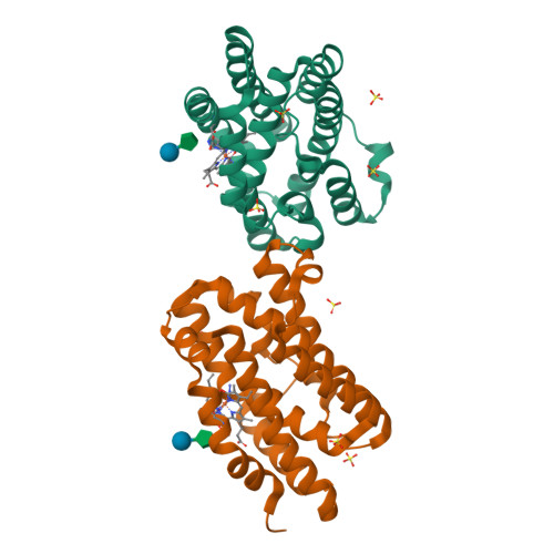
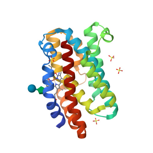
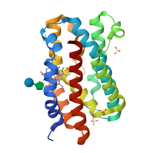
    3MOO

  • PubMed Abstract: 

    The least understood mechanism during heme degradation by the enzyme heme oxygenase (HO) is the third step of ring opening of verdoheme to biliverdin, a process which maintains iron homeostasis. In response to this mechanistic uncertainty, we launched a combined study of X-ray crystallography and theoretical QM/MM calculations, designed to elucidate the mechanism. The air-sensitive ferrous verdoheme complex of HmuO, a heme oxygenase from Corynebacterium diphtheriae, was crystallized under anaerobic conditions. Spectral analysis of the azide-bound verdoheme-HmuO complex crystals assures that the verdoheme group remains intact during the crystallization and X-ray diffraction measurement. The structure offers the first solid evidence for the presence of a water cluster in the distal pocket of this catalytically critical intermediate. The subsequent QM/MM calculations based on this crystal structure explore the reaction mechanisms starting from the FeOOH-verdoheme and FeHOOH-verdoheme complexes, which mimic, respectively, the O(2)- and H(2)O(2)-supported degradations. In both mechanisms, the rate-determining step is the initial O-O bond breaking step, which is either homolytic (for FeHOOH-verdoheme) or coupled to electron and proton transfers (in FeOOH-verdoheme). Additionally, the calculations indicate that the FeHOOH-verdoheme complex is more reactive than the FeOOH-verdoheme complex in accord with experimental findings. QM energies with embedded MM charges are close to and yield the same conclusions as full QM/MM energies. Finally, the calculations highlight the dominant influence of the distal water cluster which acts as a biocatalyst for the conversion of verdoheme to biliverdin in the two processes, by fixing the departing OH and directing it to the requisite site of attack, and by acting as a proton shuttle and a haven for the highly reactive OH(-) nucleophile.


  • Organizational Affiliation
    • Institute of Chemistry and The Lise Meitner-Minerva Center for Computational Quantum Chemistry, The Hebrew University of Jerusalem, 91904 Jerusalem, Israel.

Macromolecules
Find similar proteins by:  (by identity cutoff)  |  3D Structure
Entity ID: 1
MoleculeChains Sequence LengthOrganismDetailsImage
Heme oxygenase
A, B
215Corynebacterium diphtheriaeMutation(s): 0 
EC: 1.14.14.18
UniProt
Find proteins for Q54AI1 (Corynebacterium diphtheriae)
Explore Q54AI1 
Go to UniProtKB:  Q54AI1
Entity Groups  
Sequence Clusters30% Identity50% Identity70% Identity90% Identity95% Identity100% Identity
UniProt GroupQ54AI1
Sequence Annotations
Expand
  • Reference Sequence
Oligosaccharides

Help

Entity ID: 2
MoleculeChains Length2D Diagram Glycosylation3D Interactions
beta-D-fructofuranose-(2-1)-alpha-D-glucopyranose
C, D
2N/A
Glycosylation Resources
GlyTouCan:  G05551OP
GlyCosmos:  G05551OP
Small Molecules
Ligands 3 Unique
IDChains Name / Formula / InChI Key2D Diagram3D Interactions
VEA
Query on VEA

Download Ideal Coordinates CCD File 
K [auth A],
R [auth B]
5-OXA-PROTOPORPHYRIN IX CONTAINING FE
C33 H31 Fe N4 O5
OCHHJFVQYXRHAA-HPQJSUICSA-M
SO4
Query on SO4

Download Ideal Coordinates CCD File 
F [auth A]
G [auth A]
H [auth A]
I [auth A]
J [auth A]
SULFATE ION
O4 S
QAOWNCQODCNURD-UHFFFAOYSA-L
AZI
Query on AZI

Download Ideal Coordinates CCD File 
E [auth A],
L [auth B]
AZIDE ION
N3
IVRMZWNICZWHMI-UHFFFAOYSA-N
Biologically Interesting Molecules (External Reference) 1 Unique
Experimental Data & Validation

Experimental Data

  • Method: X-RAY DIFFRACTION
  • Resolution: 1.71 Å
  • R-Value Free:  0.213 (Depositor), 0.210 (DCC) 
  • R-Value Work:  0.178 (Depositor), 0.180 (DCC) 
  • R-Value Observed: 0.182 (Depositor) 
Space Group: C 1 2 1
Unit Cell:
Length ( Å )Angle ( ˚ )
a = 106.609α = 90
b = 63.649β = 130.34
c = 78.52γ = 90
Software Package:
Software NamePurpose
REFMACrefinement

Structure Validation

View Full Validation Report


Ligand Structure Quality Assessment 

Created with Raphaël 2.3.0Worse 01 BetterLigand structure goodness of fit to experimental dataBest fitted VEAClick on this verticalbar to view details

Entry History 

Deposition Data

Revision History  (Full details and data files)

  • Version 1.0: 2011-03-09
    Type: Initial release
  • Version 1.1: 2011-07-13
    Changes: Version format compliance
  • Version 2.0: 2020-07-29
    Type: Remediation
    Reason: Carbohydrate remediation
    Changes: Advisory, Atomic model, Data collection, Derived calculations, Non-polymer description, Structure summary
  • Version 2.1: 2023-11-01
    Changes: Data collection, Database references, Refinement description, Structure summary