3N63

Structure of neuronal nitric oxide synthase D597N/M336V mutant heme domain in complex with 6,6'-(2,2'-(5-amino-1,3-phenylene)bis(ethane-2,1-diyl))bis(4-methylpyridin-2-amine)


Experimental Data Snapshot

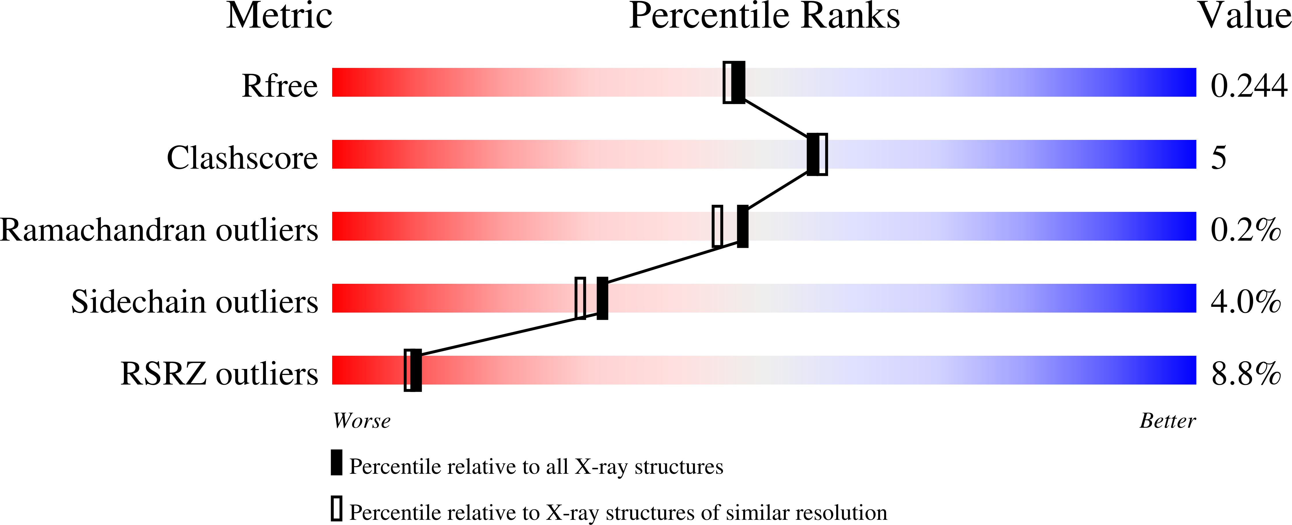
  • Method: X-RAY DIFFRACTION
  • Resolution: 2.00 Å
  • R-Value Free: 0.236 
  • R-Value Work: 0.195 
  • R-Value Observed: 0.197 

Starting Model: experimental
View more details

wwPDB Validation   3D Report Full Report


Ligand Structure Quality Assessment 


This is version 1.4 of the entry. See complete history


Literature

Role of zinc in isoform-selective inhibitor binding to neuronal nitric oxide synthase .

Delker, S.L.Xue, F.Li, H.Jamal, J.Silverman, R.B.Poulos, T.L.

(2010) Biochemistry 49: 10803-10810

  • DOI: https://doi.org/10.1021/bi1013479

  • PubMed Abstract: 

    In previous studies [Delker, S. L., et al. (2010), J. Am. Chem. Soc. 132, 5437-5442], we determined the crystal structures of neuronal nitric oxide synthase (nNOS) in complex with nNOS-selective chiral pyrrolidine inhibitors, designed to have an aminopyridine group bound over the heme where it can electrostatically interact with the conserved active site Glu residue. However, in addition to the expected binding mode with the (S,S)-cis inhibitors, an unexpected "flipped" orientation was observed for the (R,R)-cis enantiomers. In the flipped mode, the aminopyridine extends out of the active site where it interacts with one heme propionate. This prompted us to design and synthesize symmetric "double-headed" inhibitors with an aminopyridine at each end of a bridging ring structure [Xue, F., Delker, S. L., Li, H., Fang, J., Jamal, J., Martásek, P., Roman, L. J., Poulos, T. L., and Silverman, R. B. Symmetric double-headed aminopyridines, a novel strategy for potent and membrane-permeable inhibitors of neuronal nitric oxide synthase. J. Med. Chem. (submitted for publication)]. One aminopyridine should interact with the active site Glu and the other with the heme propionate. Crystal structures of these double-headed aminopyridine inhibitors in complexes with nNOS show unexpected and significant protein and heme conformational changes induced by inhibitor binding that result in removal of the tetrahydrobiopterin (H(4)B) cofactor and creation of a new Zn(2+) site. These changes are due to binding of a second inhibitor molecule that results in the displacement of H(4)B and the placement of the inhibitor pyridine group in position to serve as a Zn(2+) ligand together with Asp, His, and a chloride ion. Binding of the second inhibitor molecule and generation of the Zn(2+) site do not occur in eNOS. Structural requirements for creation of the new Zn(2+) site in nNOS were analyzed in detail. These observations open the way for the potential design of novel inhibitors selective for nNOS.


  • Organizational Affiliation

    Department of Molecular Biology and Biochemistry, University of California, Irvine, California 92697-3900, United States.


Macromolecules
Find similar proteins by:  (by identity cutoff)  |  3D Structure
Entity ID: 1
MoleculeChains Sequence LengthOrganismDetailsImage
Nitric oxide synthase
A, B
422Rattus norvegicusMutation(s): 2 
Gene Names: NOS3
EC: 1.14.13.39
UniProt
Find proteins for P29476 (Rattus norvegicus)
Explore P29476 
Go to UniProtKB:  P29476
Entity Groups  
Sequence Clusters30% Identity50% Identity70% Identity90% Identity95% Identity100% Identity
UniProt GroupP29476
Sequence Annotations
Expand
  • Reference Sequence
Small Molecules
Ligands 5 Unique
IDChains Name / Formula / InChI Key2D Diagram3D Interactions
HEM
Query on HEM

Download Ideal Coordinates CCD File 
C [auth A],
K [auth B]
PROTOPORPHYRIN IX CONTAINING FE
C34 H32 Fe N4 O4
KABFMIBPWCXCRK-RGGAHWMASA-L
XFN
Query on XFN

Download Ideal Coordinates CCD File 
D [auth A],
L [auth B]
6,6'-[(5-aminobenzene-1,3-diyl)diethane-2,1-diyl]bis(4-methylpyridin-2-amine)
C22 H27 N5
WZQDEYWNUPXXDO-UHFFFAOYSA-N
ZN
Query on ZN

Download Ideal Coordinates CCD File 
F [auth A],
G [auth A],
J [auth A]
ZINC ION
Zn
PTFCDOFLOPIGGS-UHFFFAOYSA-N
ACT
Query on ACT

Download Ideal Coordinates CCD File 
E [auth A],
M [auth B]
ACETATE ION
C2 H3 O2
QTBSBXVTEAMEQO-UHFFFAOYSA-M
CL
Query on CL

Download Ideal Coordinates CCD File 
H [auth A],
I [auth A],
N [auth B],
O [auth B]
CHLORIDE ION
Cl
VEXZGXHMUGYJMC-UHFFFAOYSA-M
Experimental Data & Validation

Experimental Data

  • Method: X-RAY DIFFRACTION
  • Resolution: 2.00 Å
  • R-Value Free: 0.236 
  • R-Value Work: 0.195 
  • R-Value Observed: 0.197 
  • Space Group: P 21 21 21
Unit Cell:
Length ( Å )Angle ( ˚ )
a = 51.802α = 90
b = 110.981β = 90
c = 163.996γ = 90
Software Package:
Software NamePurpose
REFMACrefinement
CNSrefinement
Blu-Icedata collection
DENZOdata reduction
SCALEPACKdata scaling
CNSphasing

Structure Validation

View Full Validation Report


Ligand Structure Quality Assessment 


Entry History 

Deposition Data

Revision History  (Full details and data files)

  • Version 1.0: 2011-02-09
    Type: Initial release
  • Version 1.1: 2011-07-13
    Changes: Version format compliance
  • Version 1.2: 2017-11-08
    Changes: Refinement description
  • Version 1.3: 2019-07-17
    Changes: Data collection, Refinement description
  • Version 1.4: 2023-09-06
    Changes: Data collection, Database references, Derived calculations, Refinement description